Spinal Level Identification

This Hologic DXA shows either 6 lumbar vertebral bodies or a spine where T12 has no ribs.
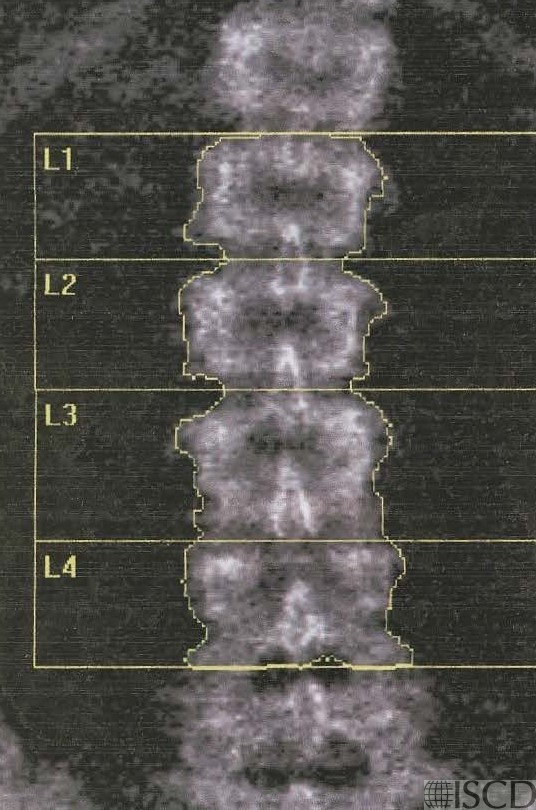
There may be 6 lumbar vertebral bodies, or T12 may not have ribs. The L4-L5 interspace is placed using the position of the iliac crest. The L5 vertebral body is the characteristic I or dog bone shape. In DXA scanning, the lumbar levels are designated from the bottom up.
Sarah L Morgan, MD, RD, CCD, The University of Alabama at Birmingham
• Peel, N.F., et al., Impact of anomalous vertebral segmentation on measurements of bone mineral density. J Bone Miner Res, 1993. 8(6): p. 719-23.
• Choplin, R.H., L. Lenchik, and S. Wuertzer, A practical approach to interpretation of Dual-Energy X-ray Absorptiometry (DXA) for assessment of bone density. . Curr Radiol Rep 2014. 2(48).
• Lewiecki, E.M. and N.E. Lane, Common mistakes in the clinical use of bone mineral density testing. Nat Clin Pract Rheumatol, 2008. 4(12): p. 667-74.
• Guan, W., et al., Lumbar Vertebrae Morphological Analysis and an Additional Approach for Vertebrae Identification in Lumbar Spine DXA Images. J Clin Densitom, 2018.
• Sarathi, V., C.V. Rakesh, and S. Tirupati, Effect of T12 Mislabeling as L1 on the Diagnosis of Low BMD at Lumbar Spine. J Clin Densitom, 2020.