Sickle Cell Anemia

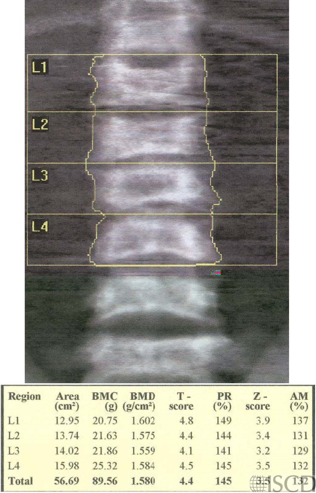
The Lincoln Log defect (H-shaped vertebrae with sharply delimited central endplate depression) or fish mouth vertebrae is seen on the spine DXA image.
This is hte hip image of the same patient showing elevated bone mineral density.
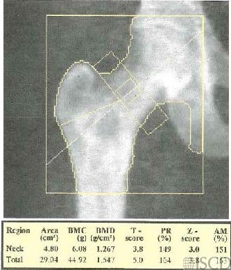
The hip and spine DXAs of a 27-year-old male patient with known sickle cell disease. Bone issues in sickle cell disease can include vaso-occlusive crisis, osteomyelitis, stress fractures, vertebral collapse, and bone marrow necrosis. In this case, the bone mineral density is elevated. It is also important to consider that hepatic iron overload in the liver, which may overlie vertebral bodies, can affect bone mineral density.
Sarah L Morgan, MD, RD, CCD, The University of Alabama at Birmingham
• Nelson, D.A., et al., Trabecular and integral bone density in adults with sickle cell disease. J Clin Densitom, 2003. 6(2): p. 125-9.
• Allard, H.M., et al., Vertebral Bone Density Measurements by DXA are Influenced by Hepatic Iron Overload in Patients with Hemoglobinopathies. J Clin Densitom, 2019. 22(3): p. 329-337.
• Almeida, A. and I. Roberts, Bone involvement in sickle cell disease. Br J Haematol, 2005. 129(4): p. 482-90