
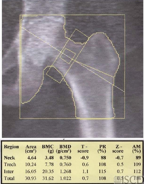
This is the baseline Hologic left hip scan.
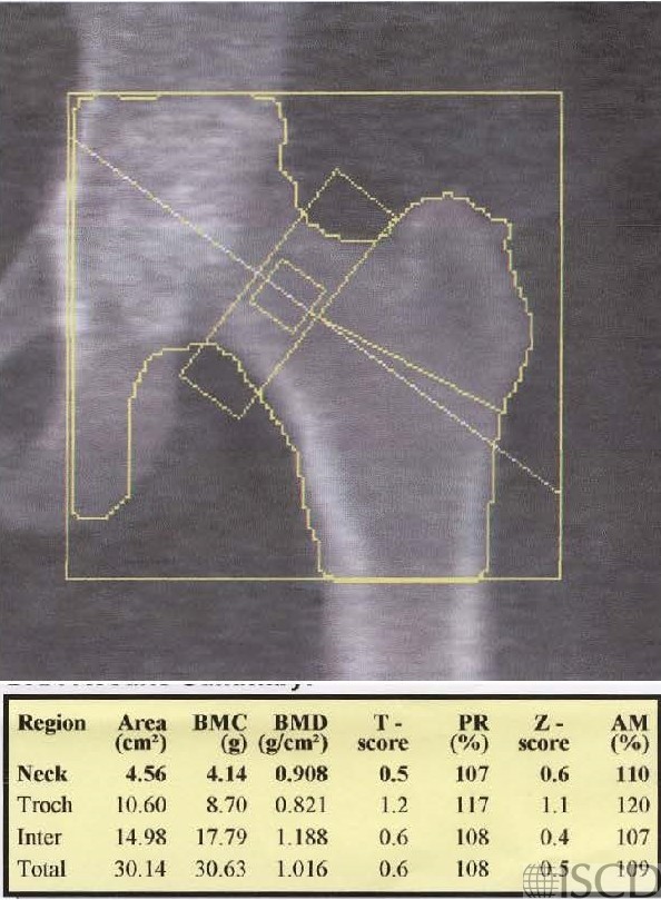
This is the follow up Hologic left hip scan 2 years later. There is a large increase at the femoral neck and no significant change a the total hip
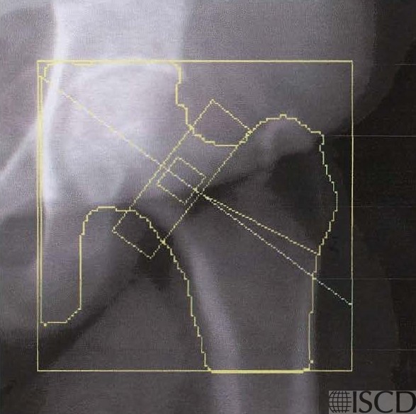
The baseline single energy view demonstrates the position of the panniculus.

The follow-up single energy view demonstrates that the panniculus is in a different position on the follow-up scan.
Case Description:
This is patient is 61″ tall and weight 273 pounds, with BMI = 51.8. The interval change was discordant between the total hip and femoral neck after 2 years. There was not a significant change at the total hip using the facility 95% confidence interval. There is a 21% increase at the femoral neck, which was a significant increase. The position of the panniculus was different between the two scans, and the position of the panniculus is documented on the single energy images. It is important to have a standard operating procedure which recommends that the patient should retract their panniculus at each scan, making the position of the panniculus consistent.
Credit:
Sarah L Morgan, MD, RD, CCD, The University of Alabama at Birmingham
References:
• Binkley, N., D. Krueger, and N. Vallarta-Ast, An overlying fat panniculus affects femur bone mass measurement. J Clin Densitom, 2003. 6(3): p. 199-204.
• Blake, G.M. and I. Fogelman, The effect of accuracy errors on the clinical interpretation of DXA scans J Clin Densitom, 2008. 11(3): p. 454.
• Fogelholm, G.M., et al., Bone mineral density during reduction, maintenance and regain of body weight in premenopausal, obese women. Osteoporos Int, 2001. 12(3): p. 199-206.
• Knapp, K.M., et al., Obesity increases precision errors in dual-energy X-ray absorptiometry measurements. J Clin Densitom, 2012. 15(3): p. 315-9.
• Knapp, K.M., et al., Obesity increases precision errors in total body dual-energy x-ray absorptiometry measurements. J Clin Densitom, 2015. 18(2): p. 209-16.
• Rajamanohara, R., et al., The effect of weight and weight change on the long-term precision of spine and hip DXA measurements. Osteoporos Int, 2011. 22(5): p. 1503-12.
• Svendsen, O.L., et al., Impact of soft tissue on in vivo accuracy of bone mineral measurements in the spine, hip, and forearm: a human cadaver study. J Bone Miner Res, 1995. 10(6): p. 868-73.
• Tothill, P. and A. Avenell, Errors in dual-energy X-ray absorptiometry of the lumbar spine owing to fat distribution and soft tissue thickness during weight change. Br J Radiol, 1994. 67(793): p. 71-5.
• Tothill, P. and A. Avenell, Anomalies in the measurement of changes in bone mineral density of the spine by dual-energy X-ray absorptiometry. Calcif Tissue Int, 1998. 63(2): p. 126-33.
• Tothill, P. and D.W. Pye, Errors due to non-uniform distribution of fat in dual X-ray absorptiometry of the lumbar spine. Br J Radiol, 1992. 65(777): p. 807-13.
• Tothill, P., N. Weir, and J. Loveland, Errors in dual-energy X-ray scanning of the hip because of nonuniform fat distribution. J Clin Densitom, 2014. 17(1): p. 91-6.