Paget’s Disease

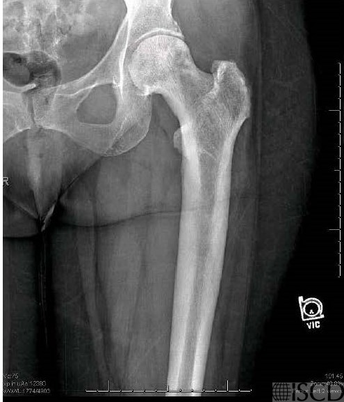
This is the left Hologic proximal femur scan of a patient with known Paget’s disease. The bone mineral density is elevated.

The corresponding radiograph in the left femur shows thickened trabeculations extending to the edge of the bone and thickened cortices extending to the middle of the shaft.
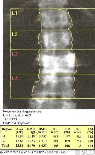
This is the lumbar spine scan of a patient with known Paget’s disease of the spine. There is diffuse sclerosis involving the anterior and posterior elements of L3 with a posterior wedge compression fracture felt to be from the Paget’s disease. It was noted that there are both sclerotic and lytic components in the L3 vertebral body. L3 and L4 are omitted from analysis.
These cases show typical findings of Paget’s disease that may seen on a DXA scan.
Sarah L Morgan, MD, RD, CCD, The University of Alabama at Birmingham
• Polyzos, S.A., et al., Dual-energy X-ray absorptiometry and quantitative ultrasound in patients with Paget’s disease of bone before and after treatment with zoledronic acid: association with serum bone markers and Dickkopf-1. J Clin Densitom, 2010. 13(2): p. 190-6.
• Cundy, T., Paget’s disease of bone. Metabolism, 2018. 80: p. 5-14.
• Tripto-Shkolnik, L. and Y. Liel, Paget’s Disease on Bone Mineral Density Examination. Qjm, 2020.
• Shanmugam, N., K. Chasse, and S.M. Gupta, Spurious elevation of bone mass secondary to Paget disease in a patient with osteoporosis. Clin Nucl Med, 2006. 31(9): p. 575-7.
• Smith, S.E., et al., From the archives of the AFIP. Radiologic spectrum of Paget disease of bone and its complications with pathologic correlation. Radiographics, 2002. 22(5): p. 1191-216.
• Vasireddy, S. and J.P. Halsey, Incidental detection of lumbar Paget’s disease by bone densitometry. Rheumatology (Oxford), 2001. 40(12): p. 1424-5.
• Wittenberg, A., The rugger jersey spine sign. Radiology, 2004. 230(2): p. 491-2