Osteogensis Imperfecta

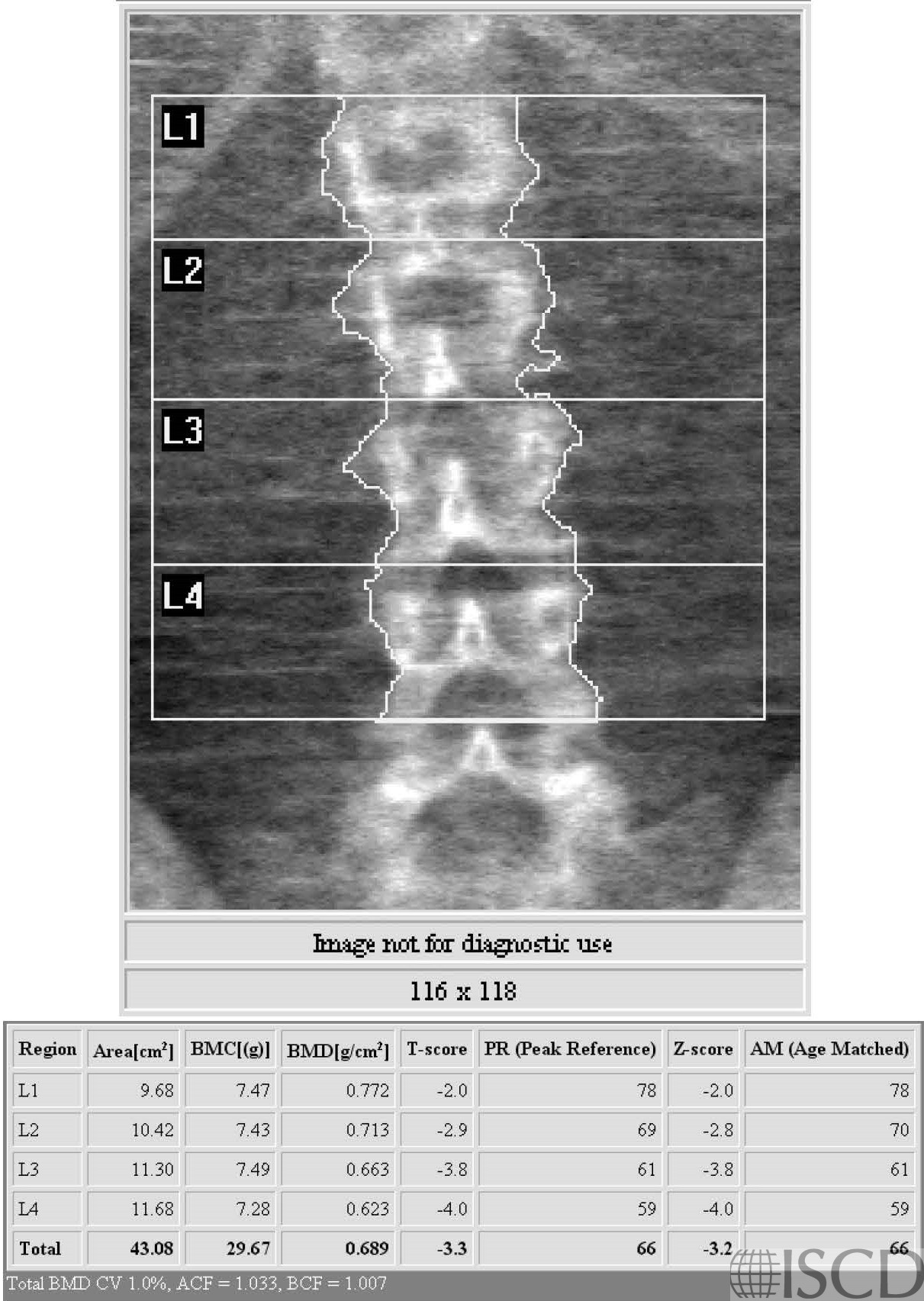
This is the Hologic spine DXA from a premenopausal female with osteogenesis imperfecta.
This is the Hologic left proximal femur scan from a premenopausal female with osteogenesis imperfecta.
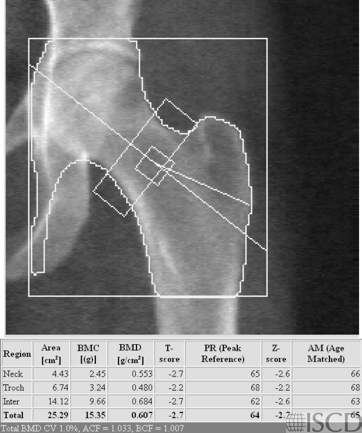
These are the scans from a 22-year-old premenopausal female referred with fractures beginning as a child. She had genetic testing and the ultimate diagnosis was osteogenesis imperfecta. It would be correct to use a Z-score in interpretation, and the diagnosis would be bone mineral density below the expected range for age.
Sarah L Morgan, MD, RD, CCD, The University of Alabama at Birmingham.
• Tournis, S. and A.D. Dede, Osteogenesis imperfecta – A clinical update. Metabolism, 2017.
• Fratzl-Zelman, N., et al., Bone mass and mineralization in osteogenesis imperfecta. Wien Med Wochenschr, 2015. 165(13-14): p. 271-7.