Normal Left Proximal Femur Scan from a GE iDXA Densitometer

Image of a Normal Left Proximal Femur DXA Scan

Densitometry BMD/T-score Reference Graph
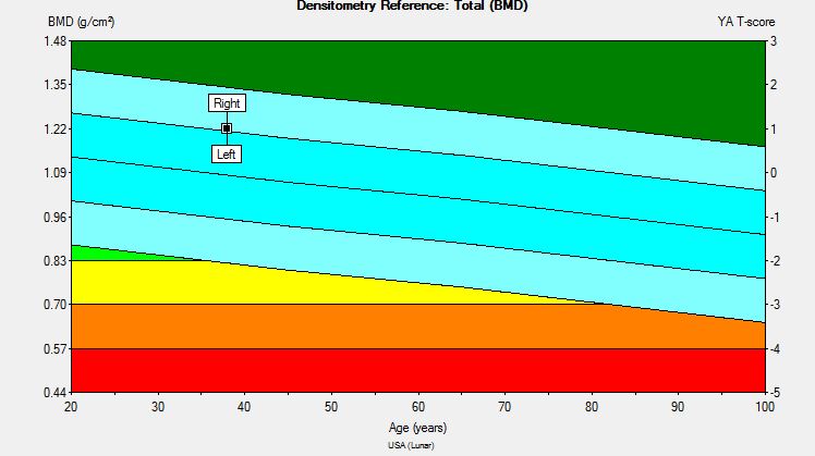
Normal left proximal femur scan from a GE iDXA densitometer from a 38-year-old, healthy, caucasian man.
Note: The shaft of the femur is straight, the hip is internally rotated using the manufacturer’s positioning triangle ensuring minimal exposure of the lesser trochanter, bone edges are correctly placed, and the total hip is the sum of the femoral neck, greater trochanter, Wards region (intertrochanteric region), and femoral shaft.
Criteria for obtaining an Optimum DXA scan using a GE densitometer:
• Correct scan mode used: thin mode (< 13cm), standard mode (13-25cm), thick mode (> 25cm)
• Sufficient pelvis and shaft separation
• Femur shaft reasonably straight (angle threshold ≤ 10 degrees)
• Proper femur rotation (lesser trochanter visibility threshold ≤ 5mm)
• Optimal contrast and brightness
• ROIs properly defined
• Tissue region and bone edges properly defined
• Results consistent with previous scan [bone area threshold (10%)]
Chris Cirnigliaro, Ph.D.
Investigator, Spinal Cord Injury Research Service
James J. Peters VA Medical Center
Lewiecki EM, Binkley N, Morgan SL, Shuhart CR, Camargos BM, Carey JJ et al. Best Practices for Dual-Energy X-ray Absorptiometry Measurement and Reporting: International Society for Clinical Densitometry Guidance. J Clin Densitom. 2016;19(2):127-40.