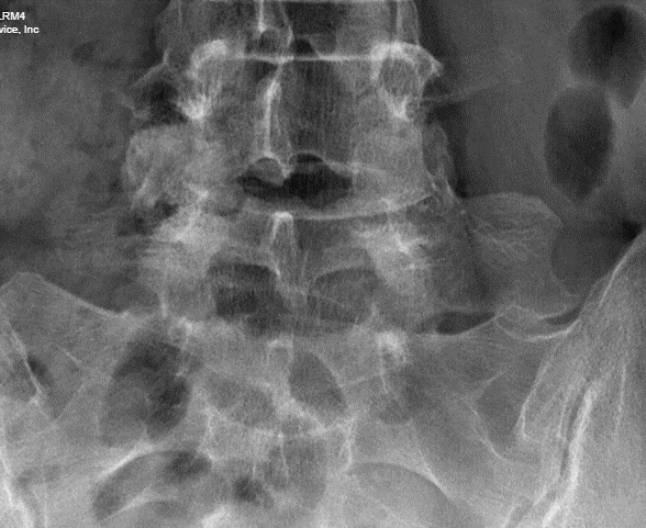
DXA scan of the lumbar spine. Hologic Horizon-A, Ver 5.3.1.2 software. Array mode. There appears to be an articulation between the left transverse process of L5 and the sacrum.

Cropped image taken from an older radiograph of the pelvic in this same patient, confirming the presence of a LSTV variant.

Case Description:
While classic anatomy convention states that there are 5 lumbar vertebra between the sacrum and the lowest thoracic (rib-bearing) vertebral body, nature has a habit of demonstrating that no rule exists without a few exceptions. Most DXA technologists are aware of situations where we encounter either four or six non rib-bearing vertebral segments on spine scanning. For consistency, the approach taught in ISCD and IOF sponsored bone densitometry courses is to count from the sacrum upwards beginning with the first segment
above the sacrum to be labelled as L5. This stems from the a study of complete spine radiographs by Peel et al (1), showing that in a group of post-menopausal women, the case of true 6 lumbar vertebra was less than 2%, with half of those presenting with a thirteenth pair of ribs at L1. giving the appearance of only 5 lumbar vertebra. More common were patients with only 11 pairs of ribs, such the labelling approach starting at the sacrum rather than at the more unreliable landmark of the lowest pair of ribs would more consistently identify and label the lumbar segments. .
We present the case of the presence of another less common segmentation variant (Image 1) often referred to as a lumbosacral transitional vertebra (LSVT) that has characteristics of both which can lead to confusion in how it and the lumbar segments above it are labeled, which affect the T-score, and Z-scores assigned to them. Sometimes LSTV’s are referred to as partially sacralization of L5, or partial lumbarization of S1, and there are various proposed mechanisms of how to classify them. (2,3).
In this case, the level was identified as L5 on DXA based on the shape of the vertebral body superior to it more representative of a typical L4 shape. Review of the patients history found an x-ray of the pelvis taken for an unrelated condition. of hip pain. No mention of a transitional vertebral articulation was mentioned in that report. A cropped section of that radiograph is presented below in Image 2.
Take home message: When reporting DXA scans, it is important to include information on the present of atypical segmentation and how you identified the lumbar vertebra. This will insure that future follow-up DXA imaging will follow the same labelling for serial measurements. In addition, it can explain potential differences in numbering with other imaging of the lumbar spine,. This could lead to the potential for the patient to undergo surgical or other interventions at an incorrect level.
We have developed a smart phrase for our reporting software that states “This patient appears to have a transitional vertebral segment. For consistency in vertebral labelling, we have chosen to label the first vertebral body superior to the sacrum as L…” The interpreter is given a drop-down list of 4,5, or 6 to fill in the ellipsis.
Credit:
Lawrence G. Jankowski, CBDT
Bone Densitometry Lab – Department of Rheumatology
Illinois Bone and Joint Institute, LLC
References:
1) Peel NF, Johnson A, Barrington NA, Smith TW, Eastell R. Impact of anomalous vertebral segmentation on measurements of bone mineral density. J Bone Miner Res. 1993 Jun;8(6):719-23. doi: 10.1002/jbmr.5650080610. PMID: 8328314.
2) Konin GP, Walz DM. Lumbosacral transitional vertebrae: classification, imaging findings, and clinical relevance. AJNR Am J Neuroradiol. 2010 Nov;31(10):1778-86. doi: 10.3174/ajnr.A2036. Epub 2010 Mar 4. PMID: 20203111; PMCID: PMC7964015.
3) Lian J, Levine N, Cho W. A review of lumbosacral transitional vertebrae and associated vertebral numeration. Eur Spine J. 2018 May;27(5):995-1004. doi: 10.1007/s00586-018-5554-8. Epub 2018 Mar 21. PMID: 29564611.
