Long Femur Scan

This image shows a Hologic left hip DXA scan. .
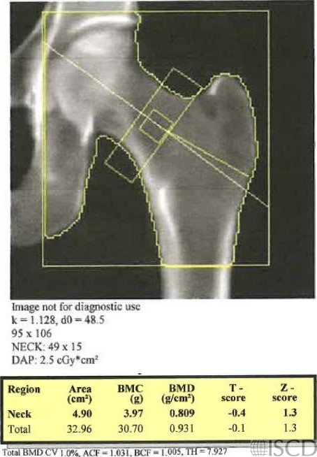
This image shows a scan with an extended length of hip scan, done on the same patient within minutes after the scan on the left. There is not a difference in bone mineral density between the nonextended and extended hip scans, using the 95% confidence intervals for the institution.

This image is the extended hip Hologic scan on the same individual. There is no evidence of a subtrochanteric fracture.
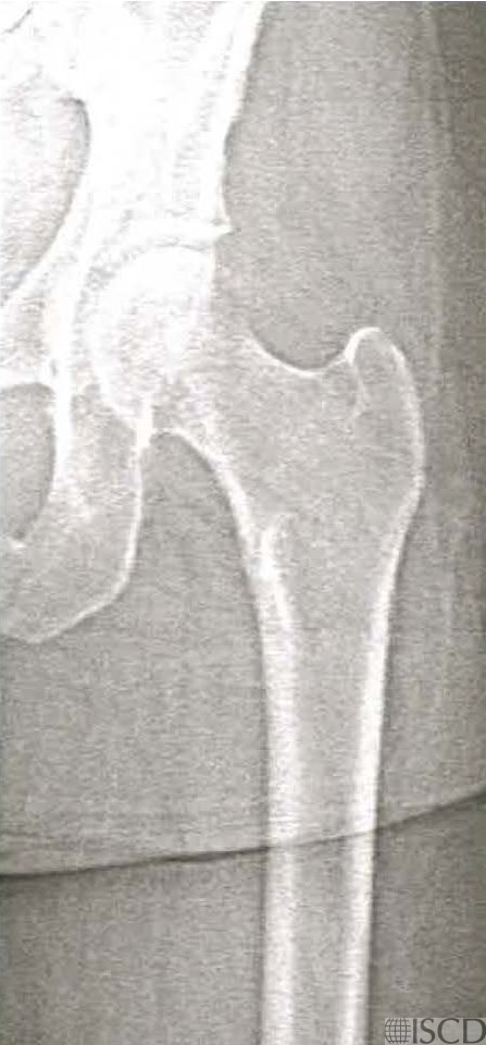
Increasing the length of the femur scan field does not affect proximal hip bone mineral density on both GE/Lunar and Hologic scanners (references below). There is an extended Hologic hip scan that is ordered to look for atypical femoral fractures.
Sarah L Morgan, MD, RD, CCD, The University of Alabama at Birmingham
• McKiernan, F.E., et al., A long femur scan field does not alter proximal femur bone mineral density measurements by dual-energy X-ray absorptiometry. J Clin Densitom, 2011. 14(3): p. 354-8.
• Prater, G.L., et al., The effect of extending femur scan length on BMD results on the Hologic Discovery-W scanner. J Clin Densitom, 2014. 17(4): p. 518-21.