Incorrect Femoral Neck Box Size


The default size of the femoral neck box on a Hologic scan is 49 x 15 pixels. The arrows point to the place in the Hologic printout which documents the size of the femoral neck box.

Case Description:

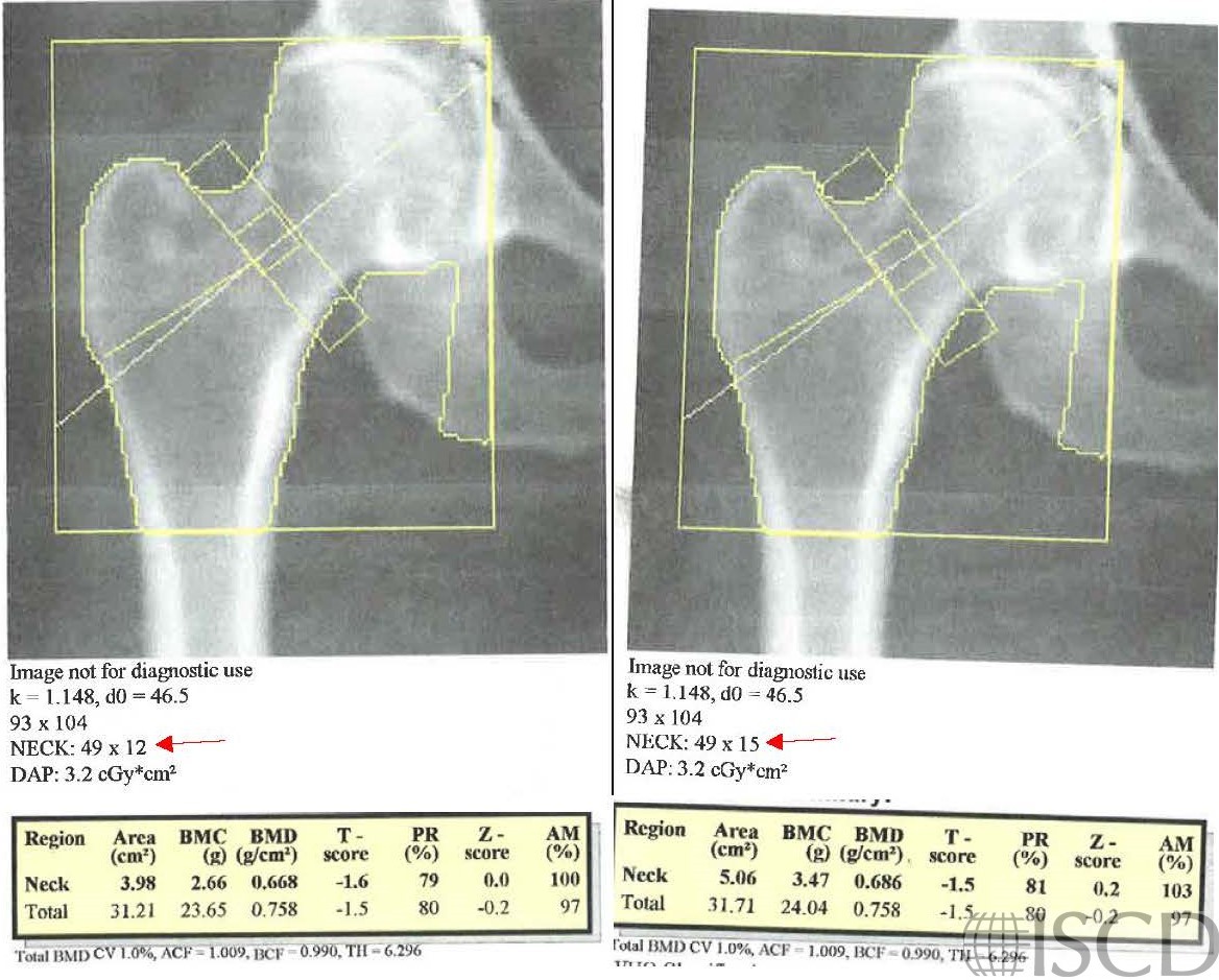
The left panel shows a Hologic hip scan with an incorrect femoral neck box size. The default is 49 x 15 pixels. The right panel shows the correct scan. For accuracy and precision, it is important to evaluate the femoral neck box positioning and size.

Credit:

Sarah L Morgan, MD, RD, CCD, The University of Alabama at Birmingham

References:
  • Watts, N.B., Fundamentals and pitfalls of bone densitometry using dual-energy X-ray absorptiometry (DXA). Osteoporos Int, 2004. 15(11): p. 847-54.
  • Choplin R.H., Lenchik L and S. Wuertzer, A practical approach to interpretation of Dual-Energy X-ray Absorptiometry (DXA) for assessment of bone density. . Curr Radiol Rep  2(48).
  • Dasher, L.G., C.D. Newton, and L. Lenchik, Dual X-ray absorptiometry in today’s clinical practice. Radiol Clin North Am, 2010. 48(3): p. 541-60.
  • Theodorou, D.J. and S.J. Theodorou, Dual-energy X-ray absorptiometry in clinical practice: application and interpretation of scans beyond the numbers. Clin Imaging, 2002. 26(1): p. 43-9.
  • Mergler, S., et al., Lumbar spine and total-body dual-energy X-ray absorptiometry in children with severe neurological impairment and intellectual disability: a pilot study of artefacts and disrupting factors. Pediatr Radiol, 2012. 42(5): p. 574-83.
  • Choi, J.S., The influence of soft tissue recognition errors on BMD value-A case report: Recipient of Young Investigator Award J Clin Densitom, 2012. 15(4): p. 483
  • Fuleihan, G.E., et al., Reproducibility of DXA absorptiometry: a model for bone loss estimates. J Bone Miner Res, 1995. 10(7): p. 1004-14.
  • Fuerst, T., et al., Quality Assurance in Bone Densitometry in Bone Densitometry and Osteoporosis  K. Genant, G. Guglielmi, and M. Jergas, Editors. 1998, Springer Berlin.
  • Hansen, K., et al., DXA Errors are Common and Likely Adversely Affect Clinical Care: DXA Quality Improvement is Needed. J Bone Miner Res 2016. 31((Suppl 1) Available at http://www.asbmr.org/ItineraryBuilder/Presentationaspx?pid=83c01c31-237b-4f07-81a5-1eeb2a7968aa&ptag=AuthorDetail&aid=00000000-0000-0000-0000-000000000000. ).
  • Promma, S., et al., Errors in Patient Positioning for Bone Mineral Density Assessment by Dual x-ray Absorptiometry: Effect of Technologist Retraining. J Clin Densitom, 2018. 21(2): p. 252-259.
  • Cetin, A., et al., Evaluation of the patient positioning during DXA measurements in daily clinical practice. Clin Rheumatol, 2008. 27(6): p. 713-5.
  • Staron, R.B., et al., Computerized bone densitometric analysis: operator-dependent errors. Radiology, 1999. 211(2): p. 467-70.
  • Baniak, N., S. Grzybowski, and W.P. Olszynski, Dual-energy x-ray absorptiometry scan autoanalysis vs manual analysis. J Clin Densitom, 2014. 17(1): p. 97-103.