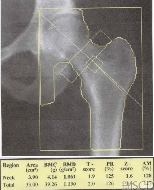
Hologic Hip Global Region of Interest Incorrect

In this Hologic left proximal femur scan, the global hp region of interest is incorrect. The bottom line, which is the most important for analysis, is down > 10 pixels from the lesser trochanter The ischium has been painted out because part of the femoral neck box overlaps the ischium.
In this image, the bottom of the hip region of interest box has been corrected. Notice that the bone mineral density of the total hip is now less because less cortical bone is included. The ischium has been painted out because a portion of the femoral neck box overlaps the ischium.
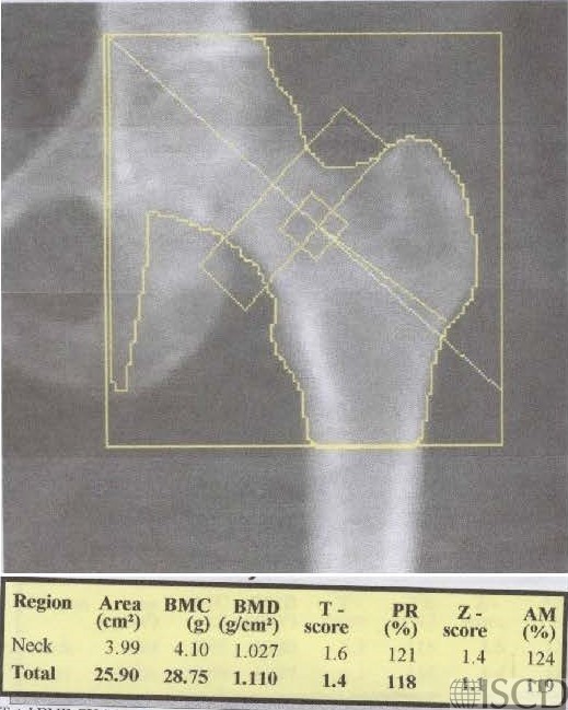
On a Hologic hip scan, the bottom of the region of interest box should be 10 pixels below the lesser trochanter. The image on the left is incorrect because the bottom of the region of interest box is >10 pixels below the lesser trochanter. By lowering the bottom of the region of interest box, the bone mineral density is generally falsely elevated, because more cortical bone is included in the total hip region of interest. The analysis on the right is the corrected analysis.
It is important to step through the entire hip analysis. Auto analysis is typically correct less than 50% of the time. The upper, inner and outer edges are all set with a blue line that touches bone and the yellow line is 5 pixels out, which places the region of interest line. The bottom line is 10 pixels below the lesser trochanter.
In both analyses, notice that a portion of the femoral neck overlaps the ischium and the ischium has been painted out.
Sarah L Morgan, MD, RD, CCD, The University of Alabama at Birmingham
• Feit, A., et al., Effect of positioning of the region of interest on bone density of the hip. . J Clin Densitom, 2020: 23(3) p 426-431.
• Morgan, S.L. and F. Peace, Do changes in the femoral neck box size make a significant difference in femoral neck BMD? . J Clin Densitom, 2011. 14 p. 156
• McKiernan, F.E., et al., A long femur scan field does not alter proximal femur bone mineral density measurements by dual-energy X-ray absorptiometry. J Clin Densitom, 2011. 14(3): p. 354-8.
• Prater, G.L., et al., The effect of extending femur scan length on BMD results on the Hologic Discovery-W scanner. J Clin Densitom, 2014. 17(4): p. 518-21.
• Celik, O., et al., The effect of hip rotation on bone mineral density of the proximal femur measured by dual energy X-ray absorptiometry. Eklem Hastalik Cerrahisi, 2009. 20(2): p. 71-7.
• Cheng, X.G., et al., Effects of anteversion on femoral bone mineral density and geometry measured by dual energy X-ray absorptiometry: a cadaver study. Bone, 1997. 21(1): p. 113-7.
• Girard, M.S., et al., Measured femoral density by dual-energy X-ray absorptiometry as a function of rotation. Orthop Rev, 1994. 23(1): p. 38-40.
• Goh, J.C., S.L. Low, and K. Bose, Effect of femoral rotation on bone mineral density measurements with dual energy X-ray absorptiometry. Calcif Tissue Int, 1995. 57(5): p. 340-3.
• Lekamwasam, S. and R.S. Lenora, Effect of leg rotation on hip bone mineral density measurements. J Clin Densitom, 2003. 6(4): p. 331-6.
• Rosenthall, L., Range of change of measured BMD in the femoral neck and total hip with rotation in women. J Bone Miner Metab, 2004. 22(5): p. 496-9.
• Tang, H., S.M. Ren, and X.Z. Luo, [Effect of femoral rotation on hip bone mineral density measurement]. Zhongguo Yi Xue Ke Xue Yuan Xue Bao, 2003. 25(3): p. 267-70.
• Wilson, C.R., et al., The effect of positioning on dual energy X-ray bone densitometry of the proximal femur. Bone Miner, 1991. 13(1): p. 69-76.
• McKiernan, F. and W. Washington, Effect of subtle positioning flaws on measured bone mineral density of the hip. J Clin Densitom, 2005. 8(3): p. 330-4.
• Hans, D., et al., Effects of a new positioner on the precision of hip bone mineral density measurements. J Bone Miner Res, 1997. 12(8): p. 1289-94.