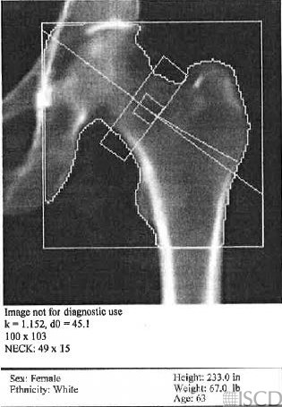
FRAX Does Not Calculate

In this Hologic hip scan the height and weight were flipped. Therefore FRAX did not calculate.
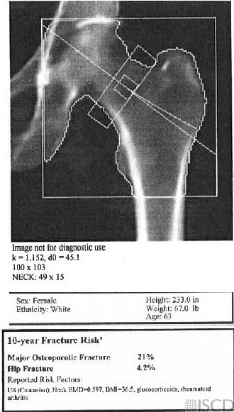
This is the corrected scan with the heigh and weight correctly entered. FRAX now calculates.
In the top panel is a Hologic hip scan. Notice in the demographics that the height and weight were switched. Therefore, FRX did not calculate.
The bottom panel shows the corrected demographic and a FRAX score is now calculated.
Sarah L Morgan, MD, RD, CCD, The University of Alabama at Birmingham
• Watts, N.B., Fundamentals and pitfalls of bone densitometry using dual-energy X-ray absorptiometry (DXA). Osteoporos Int, 2004. 15(11): p. 847-54.
• Bojinca, V., D. Opris, and M. Bonjinca, Artifacts and pitfalls in DXA scan images and interpretation. J Clin Densitom, 2012. 15(4): p. 486-487.
• Kiraç, F.S., D. Yüksel, and O.T. Yaylali, Pitfalls in the measurement of bone mineral density by the dual-energy X-ray absorptiometric method. Clin Nucl Med, 2001. 26(10): p. 874-5.
• Hansen, K., et al., DXA Errors are Common and Likely Adversely Affect Clinical Care: DXA Quality Improvement is Needed. J Bone Miner Res 2016. 31((Suppl 1) Available at http://www.asbmr.org/ItineraryBuilder/Presentation Detail.aspx?pid=83c01c31-237b-4f07-81a5-1eeb2a7968aa&ptag=AuthorDetail&aid=00000000-0000-0000-0000-000000000000. ).
• Bendavid, E.J., et al., Frequency and magnitude of analysis erros of bone density results assessed by dual x-ray absorptiometry. J Bone Miner Res, 1993. 8 (Suppl 1): p. S345.
• Binkley, N., et al., Error prevalence in DXA performance and reporting: Improving DXA quality is essential. . J Clin Densitom, 2016. ? (? ): p.? .
• El Maghraoui, A. and C. Roux, DXA scanning in clinical practice. QJM, 2008. 101(8): p. 605-17.
• Fenton, J.J., et al., Osteoporosis Overtreatment in a Regional Health Care System. JAMA Intern Med, 2016. 176(3): p. 391-3.
• Garg, M.K. and S. Kharb, Dual energy X-ray absorptiometry: Pitfalls in measurement and interpretation of bone mineral density. Indian J Endocrinol Metab, 2013. 17(2): p. 203-10.
• Kaleta, M. and S. Wronski, The most common errors in the densitometric diagnosis of osteoporosis. Ortop Traumatol Rehabil, 2001. 3(3): p. 338-44.
• Kim, T.Y. and A.L. Schafer, Variability in DXA Reporting and Other Challenges in Osteoporosis Evaluation. JAMA Intern Med, 2016. 176(3): p. 393-5.
• Krueger, D., et al., DXA Errors Are Common and Reduced by Use of a Reporting Template. J Clin Densitom, 2019. 22(1): p. 115-124.
• Lewiecki, E.M., N. Binkley, and S.M. Petak, DXA quality matters. J Clin Densitom, 2006. 9(4): p. 388-92.
• Lewiecki, E.M. and N.E. Lane, Common mistakes in the clinical use of bone mineral density testing. Nat Clin Pract Rheumatol, 2008. 4(12): p. 667-74.
• Maldonado, G., et al., Common errors in dual-energy X-ray absorptiometry scans in imaging centers in Ecuador. Arch Osteoporos, 2020. 15(1): p. 6.
• Martineau, P., M. S.L., and W.D. Leslie, Bone mineral densitometry rerporting: Pearls and pitfalls. . Can Assoc Radiol J 2020.
• Messina, C., et al., Prevalence and type of errors in dual-energy x-ray absorptiometry. Eur Radiol, 2015. 25(5): p. 1504-11.
• Lewiecki, E.M., et al., Best Practices for Dual-Energy X-ray Absorptiometry Measurement and Reporting: International Society for Clinical Densitometry Guidance. J Clin Densitom, 2016. 19(2): p. 127-40.
• El-Hajj Fuleihan, G., et al., A national random survey of bone mineral density reporting in the United States. J Clin Densitom, 2002. 5(1): p. 3-9.
• Johnston, R., et al., Quality assessment of bone density testing by DXA – Evaluation of technical and reporting deficiencies identified at a tertiary osteoporosis clinic. . J Clin Densitom, 2016. 19: p. 538-9.
• England, J.R. and P.M. Colletti, Automated Reporting of DXA Studies Using a Custom-Built Computer Program. Clin Nucl Med, 2018. 43(6): p. 474-475.
• Wachsmann, J., et al., Electronic Medical Record Integration for Streamlined DXA Reporting. J Digit Imaging, 2018. 31(2): p. 159-166.