Femoral Neck Size Different Between Baseline and Follow-up Scan


This is an outside facility scan. On the baseline scan the femoral neck box is 15 pixels wide. On the follow-up scan, the femoral neck box is much narrower and is not the Hologic default of 15 pixels wide

Case Description:

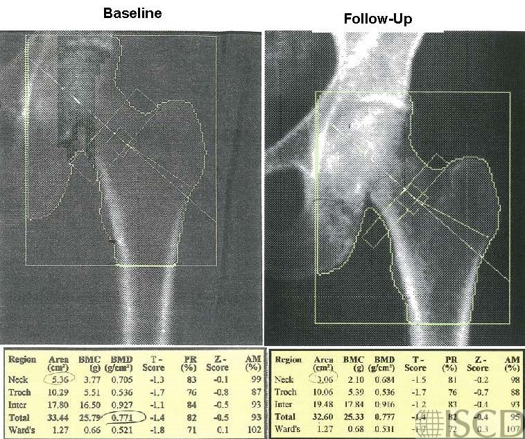
The left panel shows the baseline scan and the right panel shows the follow-up Hologic hip scan. Notice that the femoral neck box is much narrower and is not the default of 15 pixels wide on the right. This will interfere with the determination of change at the femoral neck. It is important to check and make sure that the femoral neck box is the same size on baseline and follow-up scans. This was an outside scan, so the corrected scan was not available. The patient was sent to Osteoporosis Clinic because of a concern that the femoral neck bone density was worse while on treatment. It is important to evaluate the images and technical aspects of a DXA scan.  On a Hologic scan, the size of the femoral neck box and global region of interest, in pixels, is right below the image.   In this case the sizes of the femoral neck box are obviously different.

Credit:

Sarah L Morgan, MD, RD, CCD, The University of Alabama at Birmingham

References:
  • Watts, N.B., Fundamentals and pitfalls of bone densitometry using dual-energy X-ray absorptiometry (DXA). Osteoporos Int, 2004. 15(11): p. 847-54.
  • Choplin R.H., Lenchik L and S. Wuertzer, A practical approach to interpretation of Dual-Energy X-ray Absorptiometry (DXA) for assessment of bone density. . Curr Radiol Rep  2(48).
  • Dasher, L.G., C.D. Newton, and L. Lenchik, Dual X-ray absorptiometry in today’s clinical practice. Radiol Clin North Am, 2010. 48(3): p. 541-60.
  • Theodorou, D.J. and S.J. Theodorou, Dual-energy X-ray absorptiometry in clinical practice: application and interpretation of scans beyond the numbers. Clin Imaging, 2002. 26(1): p. 43-9.
  • Mergler, S., et al., Lumbar spine and total-body dual-energy X-ray absorptiometry in children with severe neurological impairment and intellectual disability: a pilot study of artefacts and disrupting factors. Pediatr Radiol, 2012. 42(5): p. 574-83.
  • Choi, J.S., The influence of soft tissue recognition errors on BMD value-A case report: Recipient of Young Investigator Award J Clin Densitom, 2012. 15(4): p. 483
  • Fuleihan, G.E., et al., Reproducibility of DXA absorptiometry: a model for bone loss estimates. J Bone Miner Res, 1995. 10(7): p. 1004-14.
  • Fuerst, T., et al., Quality Assurance in Bone Densitometry in Bone Densitometry and Osteoporosis  K. Genant, G. Guglielmi, and M. Jergas, Editors. 1998, Springer Berlin.
  • Hansen, K., et al., DXA Errors are Common and Likely Adversely Affect Clinical Care: DXA Quality Improvement is Needed. J Bone Miner Res 2016. 31((Suppl 1) Available at http://www.asbmr.org/ItineraryBuilder/Presentationaspx?pid=83c01c31-237b-4f07-81a5-1eeb2a7968aa&ptag=AuthorDetail&aid=00000000-0000-0000-0000-000000000000. ).
  • Promma, S., et al., Errors in Patient Positioning for Bone Mineral Density Assessment by Dual x-ray Absorptiometry: Effect of Technologist Retraining. J Clin Densitom, 2018. 21(2): p. 252-259.
  • Cetin, A., et al., Evaluation of the patient positioning during DXA measurements in daily clinical practice. Clin Rheumatol, 2008. 27(6): p. 713-5.
  • Staron, R.B., et al., Computerized bone densitometric analysis: operator-dependent errors. Radiology, 1999. 211(2): p. 467-70.
  • Baniak, N., S. Grzybowski, and W.P. Olszynski, Dual-energy x-ray absorptiometry scan autoanalysis vs manual analysis. J Clin Densitom, 2014. 17(1): p. 97-103.