Femoral Neck Box Incorrectly Positioned/ Hologic

In this Hologic left hip scan,, the femoral neck is not anchored to the greater trochanter.

This is the corrected Hologic, left hip scan.
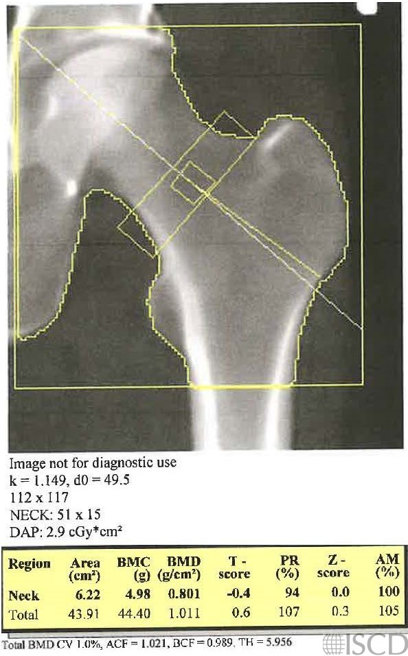
In Hologic analysis, the femoral neck should be anchored to the greater trochanter. In the image on the left, the femoral neck box is not anchored to the greater trochanter. In the corrected analysis on the right, the femoral neck is anchored to the greater trochanter. There is an artifact on the scan, but it is not within the region of interest scanned.
While changing the analysis does not make a large numerical difference in the results of the femoral neck, it is important to correctly analyze the scan. this is important not only related to accuracy, but to precision related to follow-up scanning.
On the follow-up scan the length of the femoral box is now 51 pixels, instead of the default of 49 pixels. This will not make a difference in the analysis. The important dimension is 15 pixels for the width.
Sarah L Morgan, MD, RD, CCD, The University of Alabama at Birmingham
• Feit, A., et al., Effect of positioing of the region of interest on bone density of the hip. . J Clin Densitom, 2020: 23(3) p 426-431.
*Morgan, S.L. and F. Peace, Do changes in the femoral neck box size make significant difference in femoral neck BMD? . J Clin Densitom, 2011. 14 156.