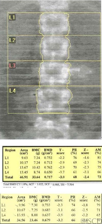
Feeding Tube


There is a feeding tube overly L3. Since it overlies bone, it would be correct to remove the L3 vertebral body from analysis. The data analysis deleting L3 is included.

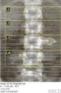
This image shows the corresponding undo view. Since the internal artifact overlies bone, that vertebral level should be omitted.

Case Description:

This Hologic lumbar spine image shows a feeding tube which overlies L3. Since it is overlying L3, it would be correct to remove L3 from the analysis.

Credit:

Sarah L Morgan, MD, RD, CCD, The University of Alabama at Birmingham

References:
  • Martineau, P., S. Bazarjani, and L.S. Zuckier, Artifacts and Incidental Findings Encountered on Dual-Energy X-Ray Absorptiometry: Atlas and Analysis. Semin Nucl Med, 2015. 45(5): p. 458-69.
  • Bazzocchi, A., et al., Incidental findings with dual-energy X-ray absorptiometry: spectrum of possible diagnoses. Calcif Tissue Int, 2012. 91(2): p. 149-56.
  • Bojinca, V., D. Opris, and M. Bonjinca, Artifacts and pitfalls in DXA scan images and interpretation. J Clin Densitom, 2012. 15(4): p. 486-487.
  • Morgan, S.L. and G.L. Prater, Quality in dual-energy X-ray absorptiometry scans. Bone, 2017.