
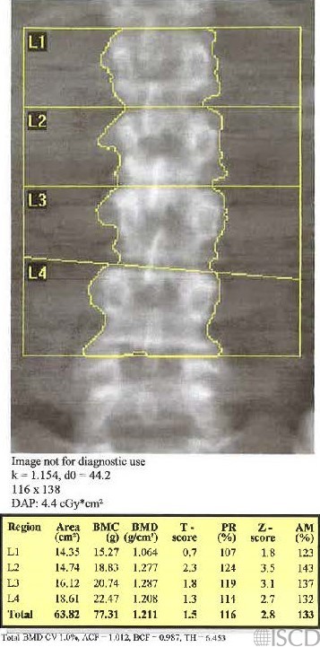
This is the baseline scan. There are degenerative changes the the Z-score is elevated (see Whyte reference).
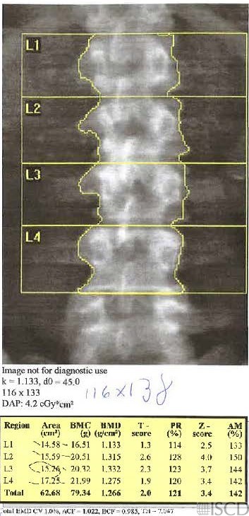
This is the follow up scan. The length of the region of interest is smaller than the baseline by 5 pixels.
Case Description:
There can be increased degenerative changes in the lumbar spine with time. This may change the size of the global region of interest at follow-up (generally the spine global region of interest is copied at follow-up). At our center, we have adopted a 3 pixel rule (not an ISCD consensus). If the global ROI size is different by >3 pixels, the new DXA scan is considered to be a new baseline. Other investigators have suggested that the corresponding areas between lumbar spine scans should generally be within 5% on follow-up scans. Since Bone Mineral Density = Bone Mineral Content/area, changes in area over time must be considered in the effect they may have on bone mineral density. On a Hologic scan, the size of the lumbar spine global region of interest may be found below the image. The default size of the width of the lumbar spine region of interest box is 116 pixels in Hologic.
Credit:
Sarah L Morgan, MD, RD, CCD, The University of Alabama at Birmingham
References:
• Kline, G.A. and D.A. Hanley, Differences of vertebral area in serial bone density measurements: a common source of potential error in interpretation of BMD change. J Clin Densitom, 2006. 9(4): p. 419-24.
• Leslie, W.D., The impact of bone area on short-term bone density precision. J Clin Densitom, 2006. 9(2): p. 150-3.
* Whyte, M.P., Misinterpretation of osteodensitometry with high bone density: BMD Z > or = + 2.5 is not “normal”. J Clin Densitom, 2005. 8(1): p. 1-6.