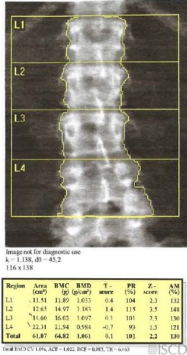
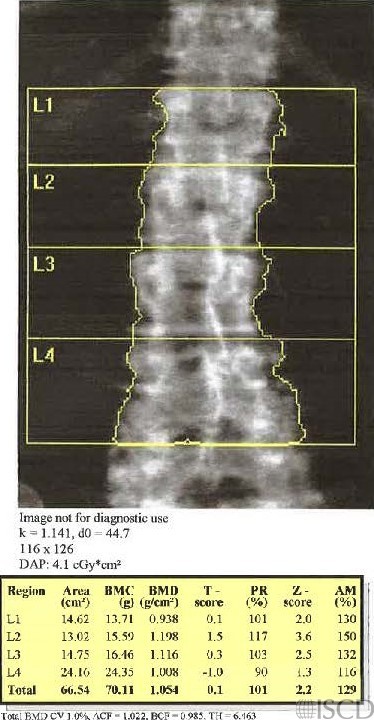
Change of Size of Lumbar Spine Region of Interest Over Time

This is the baseline scan. The size of the spine region of interest is 116 x 138 pixels
This is the follow-up scan 12 years later. There are more degenerative changes and the size of the spine global region of interest is now 116 x 126 pixels.
This is the follow up analysis showing what the analysis woud look like if the size of the ROI was 116 x 138 pixels.

There is a 12-year difference between the scan on the left and on the right panels. The initial ROI size was 116 x 138. At the 12-year follow-up, the size is 116 x 126. The bottom panel shows would the analysis would look like if the ROI size was made to be 116 x 138. There have been degenerative changes in the spine in the interim. At our institution, we have an arbitrary 3 pixel rule (not an ISCD consensus). Since the ROI size could not be brought to within 3 pixels, the new analysis is considered a new baseline. Since Bone Mineral Density = Bone Mineral Content/area, differences in the size of the region of interest could have an effect on measured BMD and also on precision.
Sarah L Morgan, MD, RD, CCD, The University of Alabama at Birmingham
• Kline, G.A. and D.A. Hanley, Differences of vertebral area in serial bone density measurements: a common source of potential error in interpretation of BMD change. J Clin Densitom, 2006. 9(4): p. 419-24.
• Leslie, W.D., The impact of bone area on short-term bone density precision. J Clin Densitom, 2006. 9(2): p. 150-3.