Catheter

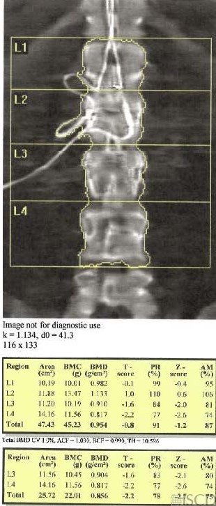
A catheter overiles l1 and L2, so those levels would be removed from analysis. The bone mineral density goes from normal to osteopenia with those levels removed from analysis.
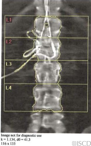
This shows the Hologic undo view of the image. There is also cathether present lateral to L3 on the right and this indicates that the artifact is removed from the soft tissue baseline. Therefore, L3 was included in the analysis.
A catheter overlies L1 and L2 in the Hologic lumbar spine image. Since the artifact overlies bone, those levels would be removed from analysis. The bone density goes fro normal to osteopenia with the removal of the overlying artifacts.
Sarah L Morgan, MD, RD, CCD, The University of Alabama at Birmingham