
Can this spine be used in diagnosis?
AP spine Density
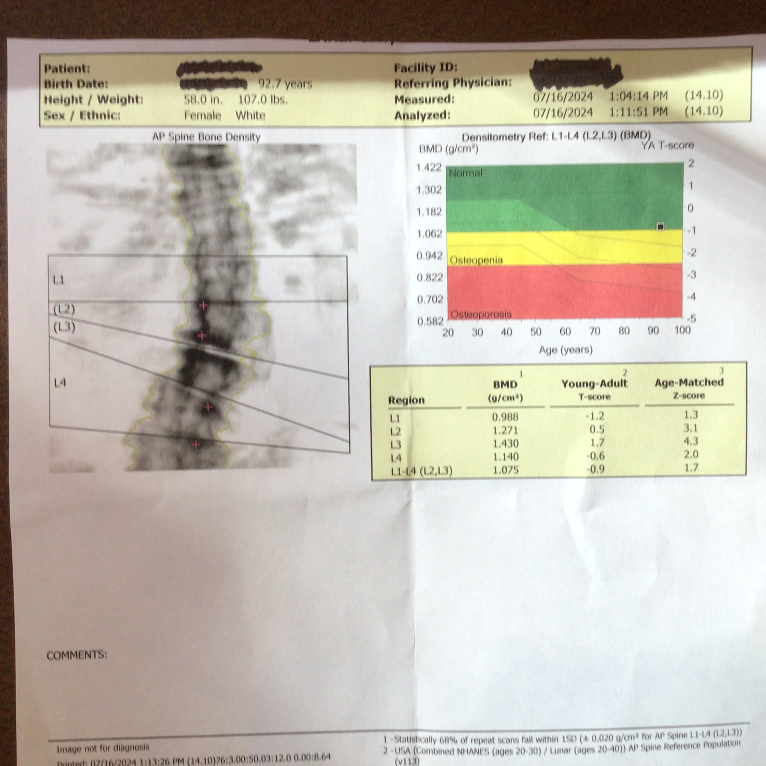
Case Description:
This is a GE Lunar Prodigy AP spine scan with L2 and L3 excluded due to differences in T-scores. Because of the degenerative changes present in the spine, this spine is not useful for diagnosis and followup. The hip site would be used and a 1/3 radius scan should be added in. There are also issues with positioning of the intervertebral marker at the bottom of the vertebral body designated as L4.
Credit:
Sheila Gatton ARRT (BD, M, R); Sutter Health, California
References:
- Watts, N.B., Fundamentals and pitfalls of bone densitometry using dual-energy X-ray absorptiometry (DXA). Osteoporos Int, 2004. 15(11): p. 847-54.
- Choplin R.H., Lenchik L and S. Wuertzer, A practical approach to interpretation of Dual-Energy X-ray Absorptiometry (DXA) for assessment of bone density. . Curr Radiol Rep 2(48).
- Dasher, L.G., C.D. Newton, and L. Lenchik, Dual X-ray absorptiometry in today’s clinical practice. Radiol Clin North Am, 2010. 48(3): p. 541-60.
- Theodorou, D.J. and S.J. Theodorou, Dual-energy X-ray absorptiometry in clinical practice: application and interpretation of scans beyond the numbers. Clin Imaging, 2002. 26(1): p. 43-9.
- Mergler, S., et al., Lumbar spine and total-body dual-energy X-ray absorptiometry in children with severe neurological impairment and intellectual disability: a pilot study of artefacts and disrupting factors. Pediatr Radiol, 2012. 42(5): p. 574-83.
- Choi, J.S., The influence of soft tissue recognition errors on BMD value-A case report: Recipient of Young Investigator Award J Clin Densitom, 2012. 15(4): p. 483
- Fuleihan, G.E., et al., Reproducibility of DXA absorptiometry: a model for bone loss estimates. J Bone Miner Res, 1995. 10(7): p. 1004-14.
- Fuerst, T., et al., Quality Assurance in Bone Densitometry in Bone Densitometry and Osteoporosis K. Genant, G. Guglielmi, and M. Jergas, Editors. 1998, Springer Berlin.
- Hansen, K., et al., DXA Errors are Common and Likely Adversely Affect Clinical Care: DXA Quality Improvement is Needed. J Bone Miner Res 2016. 31((Suppl 1) Available at http://www.asbmr.org/ItineraryBuilder/Presentationaspx?pid=83c01c31-237b-4f07-81a5-1eeb2a7968aa&ptag=AuthorDetail&aid=00000000-0000-0000-0000-000000000000. ).
- Promma, S., et al., Errors in Patient Positioning for Bone Mineral Density Assessment by Dual x-ray Absorptiometry: Effect of Technologist Retraining. J Clin Densitom, 2018. 21(2): p. 252-259.
- Cetin, A., et al., Evaluation of the patient positioning during DXA measurements in daily clinical practice. Clin Rheumatol, 2008. 27(6): p. 713-5.
- Staron, R.B., et al., Computerized bone densitometric analysis: operator-dependent errors. Radiology, 1999. 211(2): p. 467-70.
- Baniak, N., S. Grzybowski, and W.P. Olszynski, Dual-energy x-ray absorptiometry scan autoanalysis vs manual analysis. J Clin Densitom, 2014. 17(1): p. 97-103.