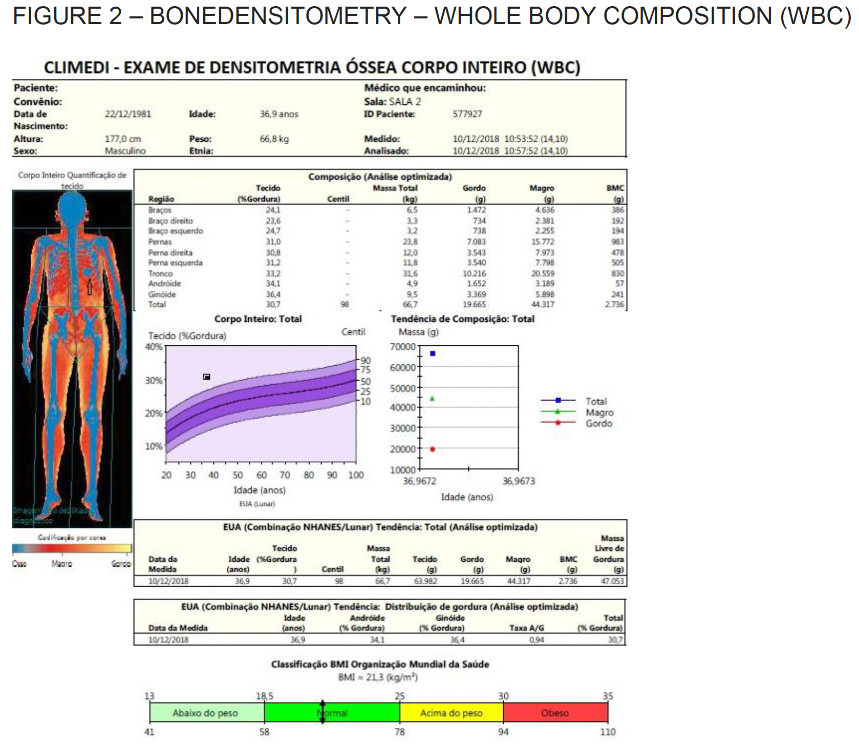
BONE DENSITOMETRY SHOWING ARTIFACT CAUSED BY CALCIFIED SPLEEN IN PATIENT WITH SICKLE CELL ANEMIA

Whole Body Composition Anterior.

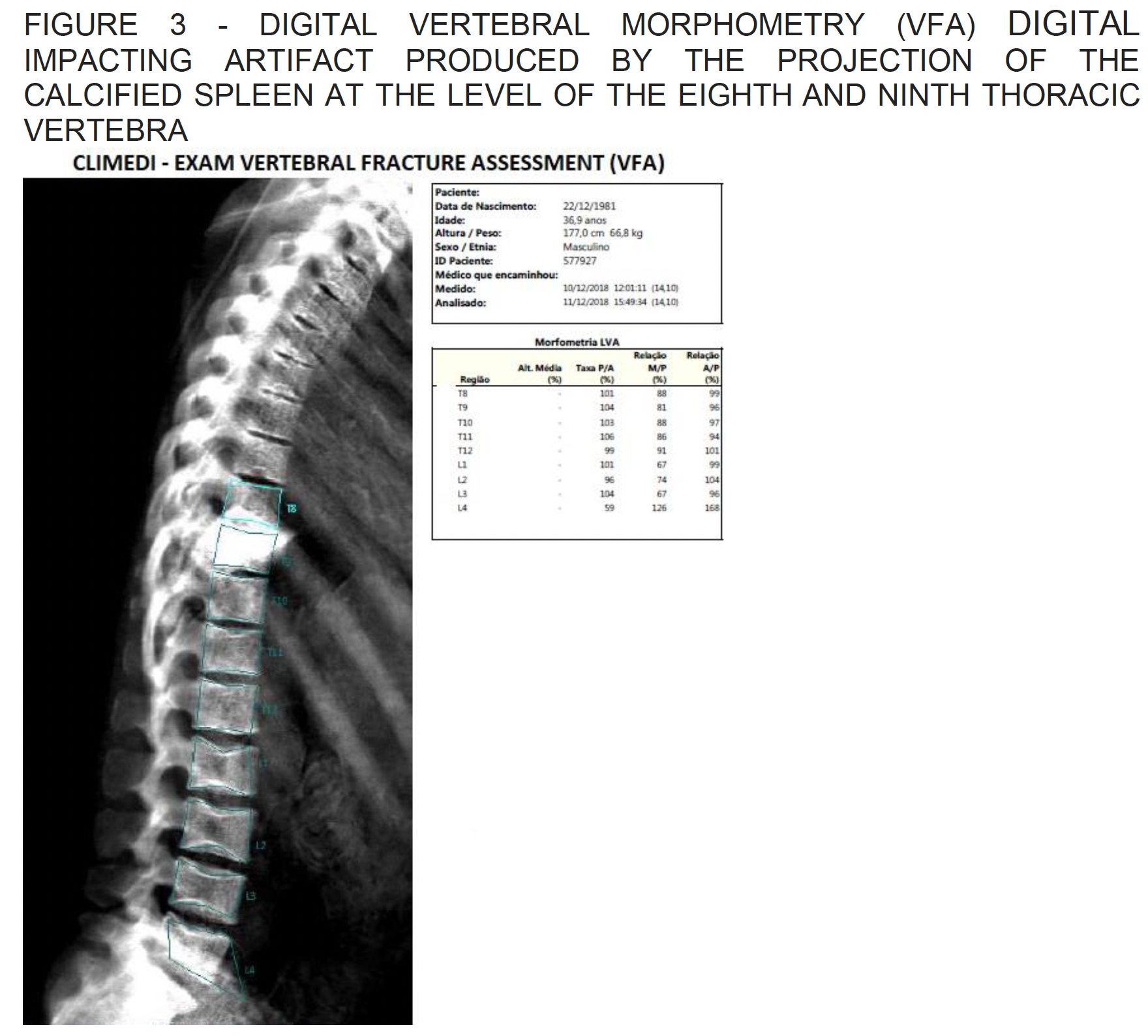
Digital Vertebral morphometry – impacting artifact produced by the projection of the calcified spleen at the level of the eighth and ninth thoracic vertebra.
Whole Body Composition Posterior.
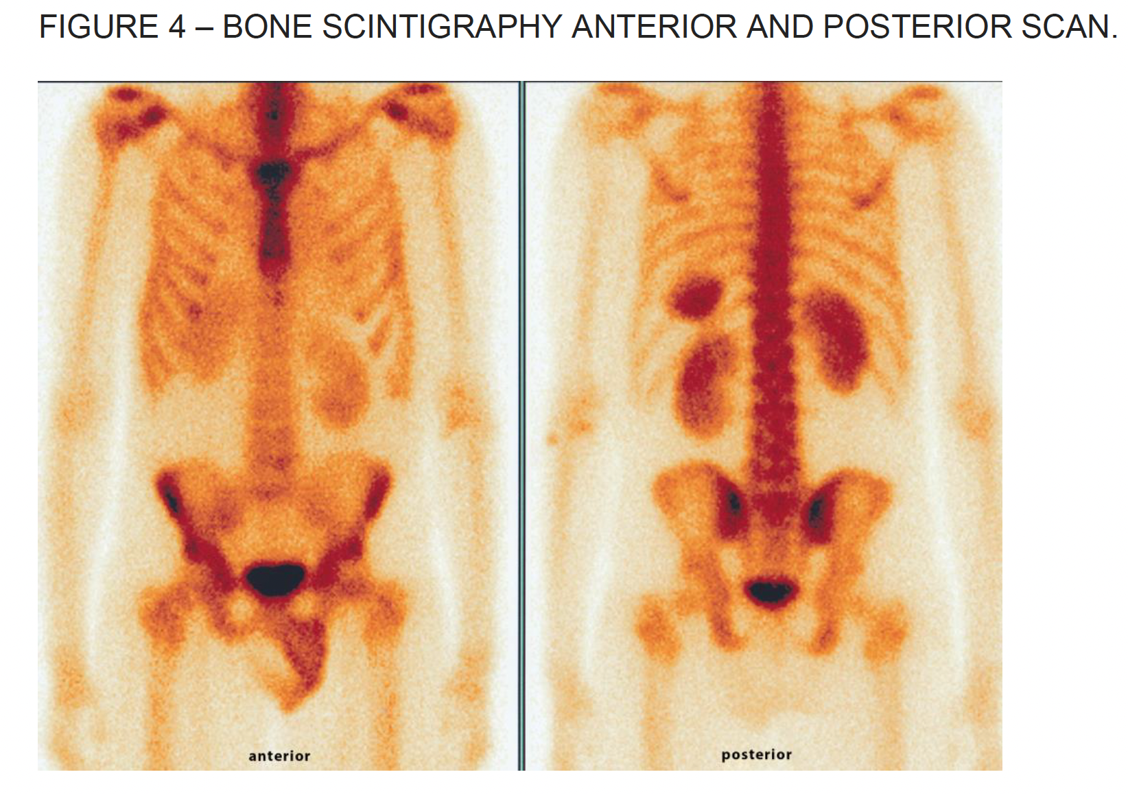
Bone Scintigraphy.
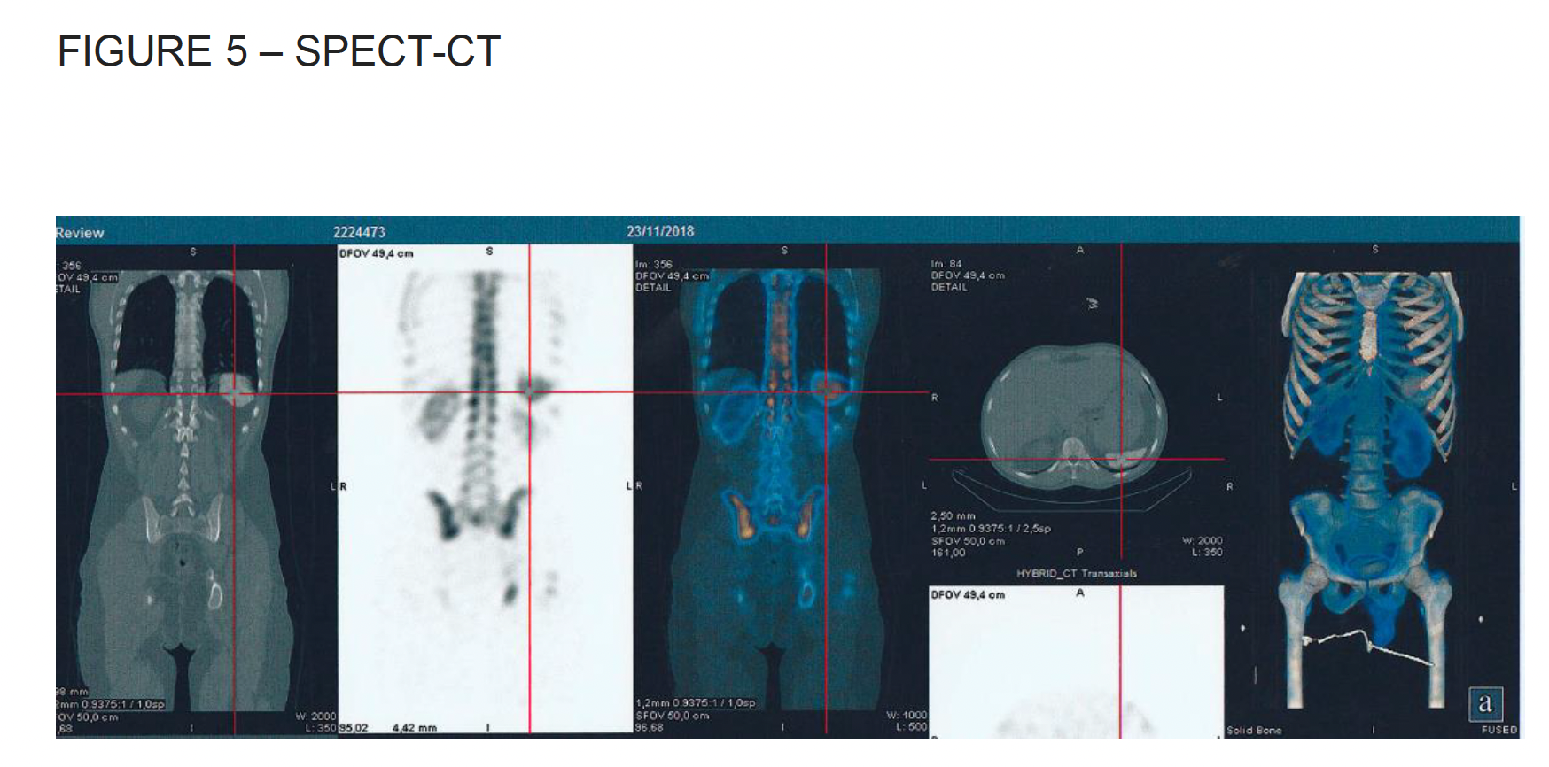
Spect-CT on bone scintigraphy
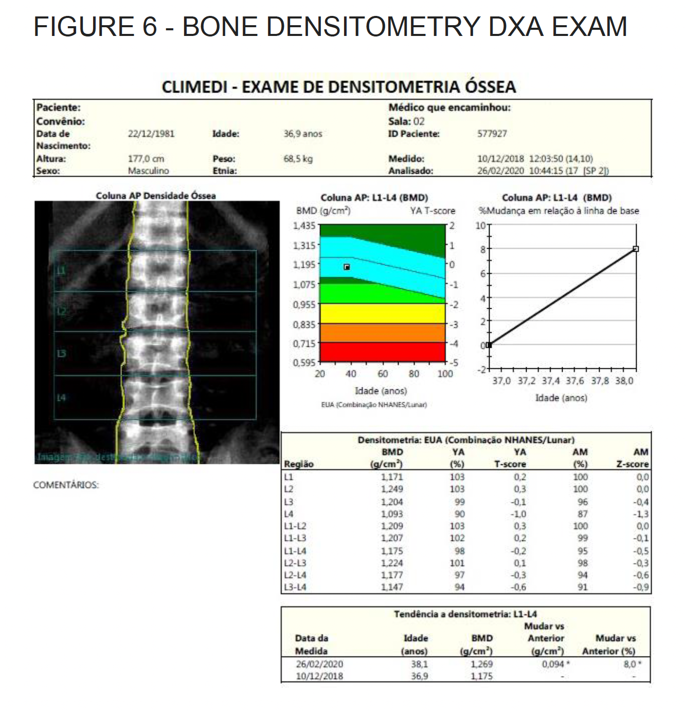
Lumbar Spine.
ABSTRACT: The purpose of this study was to present a case of incidental finding (artifact) caused by the calcified spleen in a bone densitometry scan, whole-body composition and vertebral fracture assessment of a patient with sickle cell anemia.
INTRODUCTION: Bone mineral density (BMD) measurement by dual- energy X ray absorptiometry (DXA) is the most commonly used method to assess fracture risk. DXA utilizes two different energy X-rays to calculate BMD and by comparison to a young normative database, the T-score. In 1994, the World Health Organization defined osteoporosis based on T-score changing the paradigm of the field and forever placing DXA measurements in the center of osteoporosis diagnosis. There are numerous artifacts that can alter DXA BMD measurement. It is important for providers who interpret DXA scans to be familiar with the potential effects of artifacts and how to adjust for their presence, since erroneous measurements can lead to incorrect decisions regarding treatment and follow-up. Among the artifacts found in the bone densitometry exam, we can enumerate, metal objects such as surgical clips used in vascular surgery, lead bullets that have high density, belly button rings, barite contrasts, metallic type zippers, removable artifacts such as earrings, rings, bracelets, car keys, coins and other metallic objects and even those produced by osteophytes (that may lead to an overestimated result of the densitometric exam), osteoarthritis, fractures, end-plate sclerosis, aortic calcification, pancreatic calcification, kidney stone, gallbladder stones, radiopaque contrast material, clips, facet joint syndrome. Ultimately, these artifacts or vertebrae involved should be excluded so as not to compromise the quality of the exam (01,02,03,04).
CONCLUSIONS: This artifact or incidental finding of the calcified spleen in the examination of bone densitometry, in the study of whole-body composition and digital vertebral morphometry should be included in the current literature, so that physicians involved with this diagnostic technology become aware of this artifact.
Santana AM Joao* MD and Santana AM Sara**
*CLIMEDI – Clínica de Medicina Nuclear Endocrinologia e Diabetes, Aracaju-Se Brazil.
Post Graduated Research Fellow in Endocrinology. Upstate Medical Center Hospital. State University of New York. USA.
**Specialist in Radiology and Nuclear Medicine. CBR and SBMN.
Email: joaoamsantana@uol.com.br
01 – Artifacts and Incidental Findings Encountered on Dual-Energy X-Ray Absorptiometry: Atlas and Analysis. Seminars in Nuclear Medicine 45(5):458-69 · September 2015.
02 – Nelson B. Watts et al. Fundamentals and pitfalls of bone densitometry using dual-energy X-ray absorptiometry (DXA). Osteoporosis International Nov 2004, Volume 15, Issue 11, pp 847–854.
03 – M.K. Garg and Sandeep Kharb. Dual energy X-ray absorptiometry: Pitfalls in measurement and interpretation of bone mineral density. Indian J Endocrinol Metab. 2013 Mar-Apr; 17(2) 203-210.
04 – Kerry Siminoski, MD FRCPC et al. CAR Clinical Practice Guidelines/Directives Cliniques de la CAR. Recommendations for Bone Mineral Density reporting in Canada.
05 – Alberto Ribeiro de Souza Leão e cols. Aspectos radiológicos das alterações esplênicas. Rev Imagem 2005; 27(4):253-262
06 – Gael J. Lonergan et al. Sickle Cell Anemia. 2001. https://doi.org/10.1148/radiographics.21.4.g01jl23971