Benign Bone Lesions – Bone Infarct

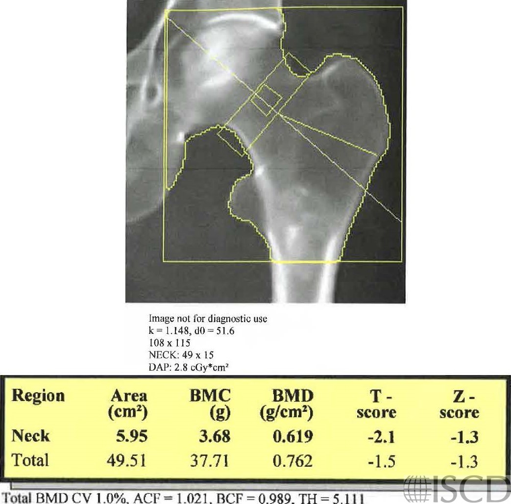
There is a rounded lesion lateral and inferior to the left lesser trochanter on this Hologic proximal hip scan.
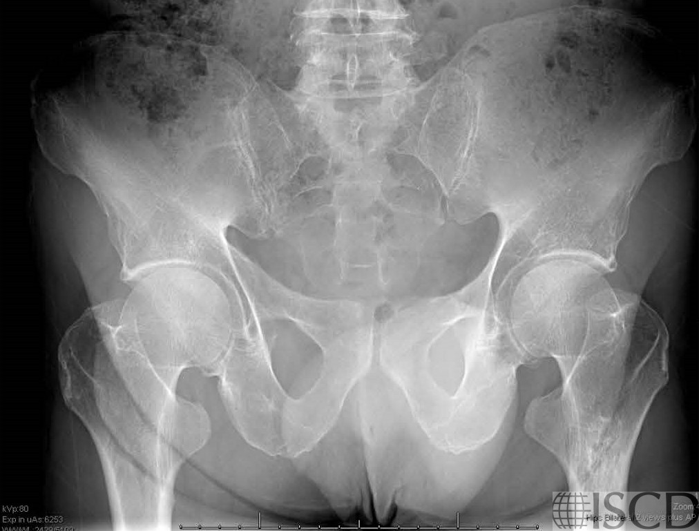
This is the accompanying radiograph. The lesion has been followed longitudinally and has been read as a bone infarct.
There is a rounded lesion lateral and inferior to the left lesser trochanter on this Hologic left proximal hip scan. The lesion has been followed longitudinally and has been read as a bone infarct on the accompanying radiograph.
Sarah L Morgan, MD, RD, CCD, The University of Alabama at Birmingham
• Martineau, P., S. Bazarjani, and L.S. Zuckier, Artifacts and Incidental Findings Encountered on Dual-Energy X-Ray Absorptiometry: Atlas and Analysis. Semin Nucl Med, 2015. 45(5): p. 458-69.
• Kim, S.K. and W.F. Barry, Jr., Bone islands. Radiology, 1968. 90(1): p. 77-8.
• Geniets, C., et al., Proceedings of the European Society of Musculoskeletal Radiology (ESSR) training module, Antwerp, 20-21.01.05. Part two: bone tumors. Benign bone lesions: characteristic imaging features. Jbr-btr, 2006. 89(5): p. 266-74.