Benign Bone Lesion – Bone Island

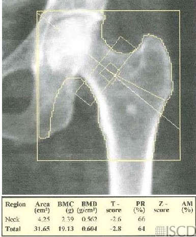
There is a bone island seen on the left Hologic proximal femur DXA scan.
The accompanying radiograph also shows the bone island.
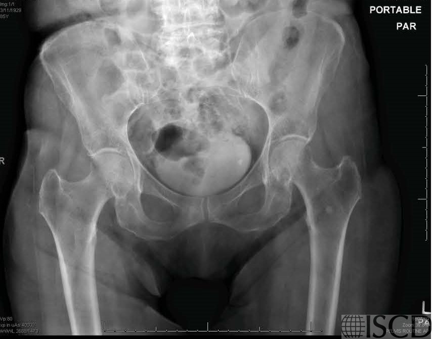
There is a bone island seen on the left Hologic proximal femur scan. On the accompanying pelvic radiograph, the sclerotic foci in the left ilium and proximal femur were read as consistent with bone islands. A bone island is also known as an enostosis and is a benign lesion. Most lesions are small and round and are composed of cortical bone within the cancellous bone.
Sarah L Morgan, MD, RD, CCD, The University of Alabama at Birmingham
• Martineau, P., S. Bazarjani, and L.S. Zuckier, Artifacts and Incidental Findings Encountered on Dual-Energy X-Ray Absorptiometry: Atlas and Analysis. Semin Nucl Med, 2015. 45(5): p. 458-69.
• Kim, S.K. and W.F. Barry, Jr., Bone islands. Radiology, 1968. 90(1): p. 77-8.
• Geniets, C., et al., Proceedings of the European Society of Musculoskeletal Radiology (ESSR) training module, Antwerp, 20-21.01.05. Part two: bone tumors. Benign bone lesions: characteristic imaging features. Jbr-btr, 2006. 89(5): p. 266-74.