Normal Hologic Hip Scan

The is an image of a normal left Hologic proximal femur scan.
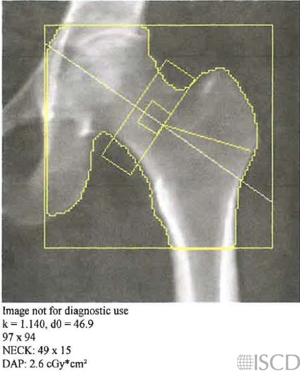
This image shows the femoral neck box and the femoral neck region of interest.

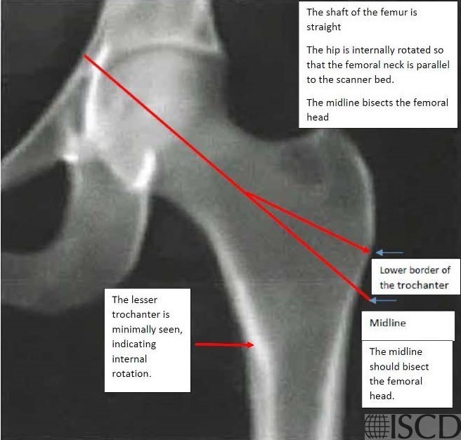
The shaft of the femur should be straight and the hip should be internally rotated.
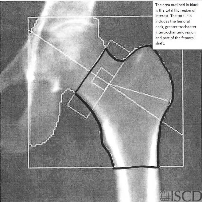
The image shows the total hip region of interest.
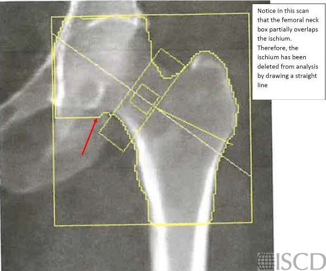
If the femoral neck overlaps the ischium, the ischium can be painted out.
These images review the placement of the regions of interest for both Hologic and GE.
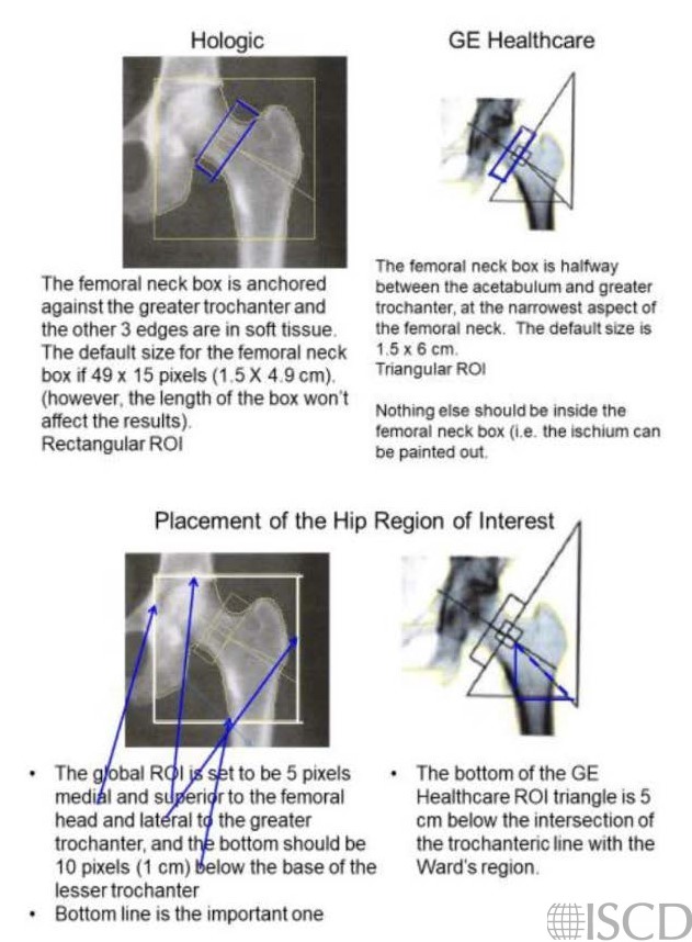
The hip that is scanned is internally rotated using a hip positioner. The hip is internally rotated because that makes the femoral neck parallel to the scanner bed. The default width of the femoral neck box is 15 pixels. The analysis is stepped through and a line is placed on various landmarks (i.e. head of the femur, greater trochanter and bottom of the lesser trochanter) and this positions the global region of interest box of the hip scan. It is the bottom of the hip global region of interest that is most important related to total hip results on a Hologic scan. The bottom of the global region of interest should be 10 pixels below the bottom of the lesser trochanter. The lesser trochanter may be better defined by altering the contrast on the scan. If the femoral neck overlaps the ischium, the edge detection may be altered to paint out that portion of the ischium.
Things to evaluate on a scan include: PARED
P – Positioning – is the patient positioned correctly
A – Artifacts – Are there artifacts in the soft tissue or over bone?
R – Region of Interest – are the regions of interest correct?
E – Edge Detection – Is the edge detection algorithm correct?
D – Databases – Are the correct databases used for the T-scores and Z-scores?
Sarah L Morgan, MD, RD, CCD, The University of Alabama at Birmingham
Feit A, Levin N, McNamara EA, Sinha P, Whittaker LG, Malabanan AO, Rosen HN. Effect of Positioning of the Region of Interest on Bone Density of the Hip. J Clin Densitom. 2020 Jul-Sep;23(3):426-431. doi: 10.1016/j.jocd.2019.04.002. Epub 2019 Apr 5. PMID: 31036446