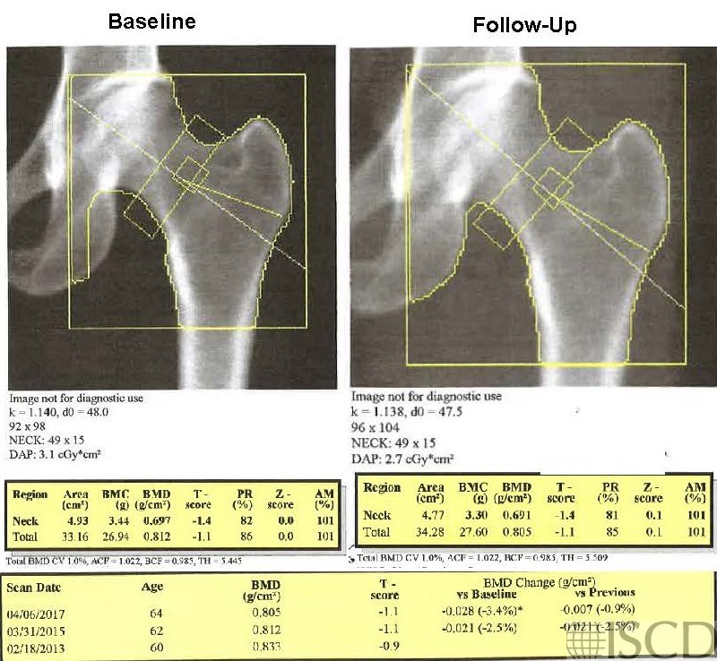
Wrong Hologic Total Hip Global Region of Interest Size on Follow-up


The upper left panel has the baseline scan. In the upper right panel the size of the hip global region of interest is not the same.  On a Hologic scan, the size of the hip global region of interest, as well as the size of the femoral neck, in pixels is below the image.  The bone map should be copied from the baseline to follow-up scan.

In this panel the global hip region of interest is now the same as baseline. The total hip bone mineral density is now less because less cortical bone from the femoral shaft is included in the total hip region of interest.  Note that the sizes of the hip global region of interest, in pixels, are the same on the baseline and follow-up scan.

Case Description:

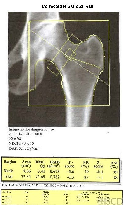
The size of the global hip region of interest is not the same in the baseline scan (upper left) and the follow-up scan (upper right). The bone map should be transferred and the size of the total hip global region of interest should be the same as on the follow-up and the baseline scan. The bottom panel shows the follow-up scan with the corrected global hip region of interest size. There is not a difference in the significance of change in this instance, using the institution 95% confidence intervals.

Credit:

Sarah L Morgan, MD, RD, CCD, The University of Alabama at Birmingham

References:
  • Watts, N.B., Fundamentals and pitfalls of bone densitometry using dual-energy X-ray absorptiometry (DXA). Osteoporos Int, 2004. 15(11): p. 847-54.
  • Choplin R.H., Lenchik L and S. Wuertzer, A practical approach to interpretation of Dual-Energy X-ray Absorptiometry (DXA) for assessment of bone density. . Curr Radiol Rep  2(48).
  • Dasher, L.G., C.D. Newton, and L. Lenchik, Dual X-ray absorptiometry in today’s clinical practice. Radiol Clin North Am, 2010. 48(3): p. 541-60.
  • Theodorou, D.J. and S.J. Theodorou, Dual-energy X-ray absorptiometry in clinical practice: application and interpretation of scans beyond the numbers. Clin Imaging, 2002. 26(1): p. 43-9.
  • Mergler, S., et al., Lumbar spine and total-body dual-energy X-ray absorptiometry in children with severe neurological impairment and intellectual disability: a pilot study of artefacts and disrupting factors. Pediatr Radiol, 2012. 42(5): p. 574-83.
  • Choi, J.S., The influence of soft tissue recognition errors on BMD value-A case report: Recipient of Young Investigator Award J Clin Densitom, 2012. 15(4): p. 483
  • Fuleihan, G.E., et al., Reproducibility of DXA absorptiometry: a model for bone loss estimates. J Bone Miner Res, 1995. 10(7): p. 1004-14.
  • Fuerst, T., et al., Quality Assurance in Bone Densitometry in Bone Densitometry and Osteoporosis  K. Genant, G. Guglielmi, and M. Jergas, Editors. 1998, Springer Berlin.
  • Hansen, K., et al., DXA Errors are Common and Likely Adversely Affect Clinical Care: DXA Quality Improvement is Needed. J Bone Miner Res 2016. 31((Suppl 1) Available at http://www.asbmr.org/ItineraryBuilder/Presentationaspx?pid=83c01c31-237b-4f07-81a5-1eeb2a7968aa&ptag=AuthorDetail&aid=00000000-0000-0000-0000-000000000000. ).
  • Promma, S., et al., Errors in Patient Positioning for Bone Mineral Density Assessment by Dual x-ray Absorptiometry: Effect of Technologist Retraining. J Clin Densitom, 2018. 21(2): p. 252-259.
  • Cetin, A., et al., Evaluation of the patient positioning during DXA measurements in daily clinical practice. Clin Rheumatol, 2008. 27(6): p. 713-5.
  • Staron, R.B., et al., Computerized bone densitometric analysis: operator-dependent errors. Radiology, 1999. 211(2): p. 467-70.
  • Baniak, N., S. Grzybowski, and W.P. Olszynski, Dual-energy x-ray absorptiometry scan autoanalysis vs manual analysis. J Clin Densitom, 2014. 17(1): p. 97-103.