Marfan Syndrome

This is the GE Healthcare DXA scan of a man with Marfan Syndrome. Additional workup demonstrated compresson fractures and L1 and L4.
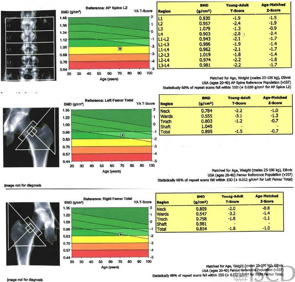
This is a GE Healthcare DXA scan from a man with known Marfan Syndrome. Compression fractures were demonstrated at L1 and L4. The literature is somewhat equivocal about wither Marfan Syndrome is associated with low bone mass.
Sarah L Morgan, MD, RD, CCD, The University of Alabama at Birmingham
• Giampietro, P.F., et al., Assessment of bone mineral density in adults and children with Marfan syndrome. Osteoporos Int, 2003. 14(7): p. 559-63.
• Giampietro, P.F., et al., Bone mineral density determinations by dual-energy x-ray absorptiometry in the management of patients with Marfan syndrome–some factors which affect the measurement. Hss j, 2007. 3(1): p. 89-92.
• Le Parc, J.M., S. Molcard, and F. Tubach, Bone mineral density in Marfan syndrome. Rheumatology (Oxford), 2001. 40(3): p. 358-9.
• Trifirò, G., et al., Areal bone mineral density in children and adolescents with Marfan syndrome: evidence of an evolving problem. Bone, 2015. 73: p. 176-80.