Hyperthyroidism

This is the Hologic DXA image from a patient with iatrogenic hyperthyroidism.

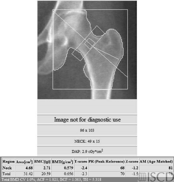
This is the Hologic left proximal femur scan from a patient with iatrogenic hyperthyroidism.
This is the baseline Hologic DXA scan from a postmenopausal woman referred to Osteoporosis Clinic with low bone mineral density. The accompanying note from the referring endocrinologist indicated that her thyroid replacement dose had been five-times the dose that was required to make her euthyroid for the past several years. Hyperthyroidism is a secondary cause of low bone mass.
Sarah L Morgan, MD, RD, CCD, The University of Alabama at Birmingham
Williams, G.R. and J.H.D. Bassett, Thyroid diseases and bone health. J Endocrinol Invest, 2018. 41(1): p. 99-109.
Segna, D., et al., Association between subclinical thyroid dysfunction and change in bone mineral density in prospective cohorts. J Intern Med, 2018. 283(1): p. 56-72.