Compression Fracture


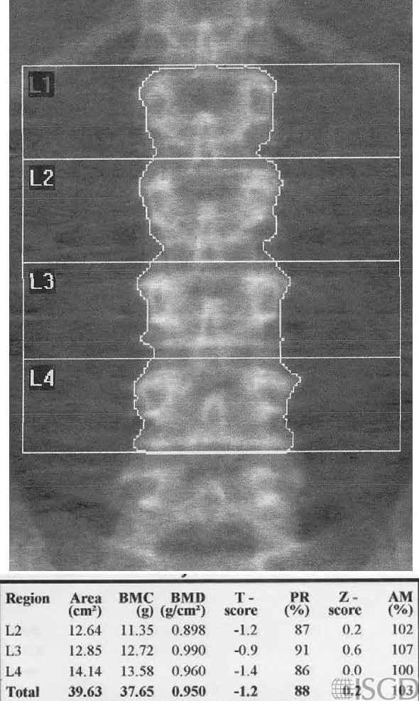
This is the baseline Hologic lumbar spine DXA scan showing no compression fractures.

The follow-up DXA scan below shows a L1 fracture.   This is accompanying lateral spine radiograph which  shows the new L1 compression fracture on follow-up.

The follow-up DXA scan after 2 years shows that the height of L1 is shortened and that there is a compression fracture. L1 is omitted from the baseline and previous DXA scan because of the presence of compression.

Case Description:

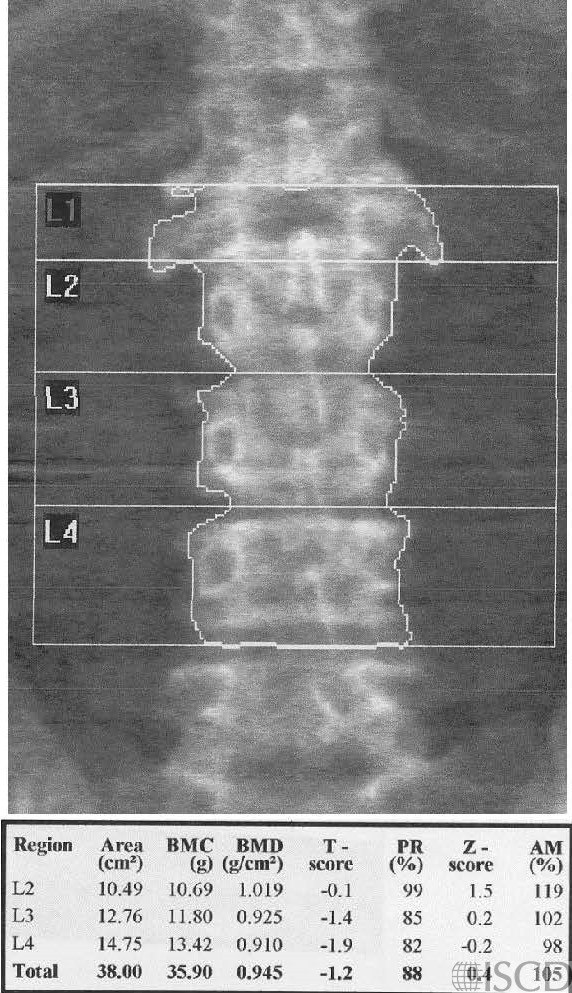
There is a baseline DXA scan showing no compression fracture and the intervening DXA scan, completed after 2 years, shows a new L1 compression fracture.

Credit:

Sarah L Morgan, MD, RD, CCD, The University of Alabama at Birmingham

References:
  • Martineau, P., S. Bazarjani, and L.S. Zuckier, Artifacts and Incidental Findings Encountered on Dual-Energy X-Ray Absorptiometry: Atlas and Analysis. Semin Nucl Med, 2015. 45(5): p. 458-69.
  • Bazzocchi, A., et al., Incidental findings with dual-energy X-ray absorptiometry: spectrum of possible diagnoses. Calcif Tissue Int, 2012. 91(2): p. 149-56
  • Lentle, B., J. Trollip, and K. Lian, The Radiology of Osteoporotic Vertebral Fractures Redux. J Clin Densitom, 2016. 19(1): p. 40-7.
  • Oei, L., et al., Osteoporotic Vertebral Fractures as Part of Systemic Disease. J Clin Densitom, 2016. 19(1): p. 70-80.
  • Ryan, P.J., et al., The effect of vertebral collapse on spinal bone mineral density measurements in osteoporosis. Bone Miner, 1992. 18(3): p. 267-72.
  • Morgan, S.L., et al., Nonprogression of vertebral area or bone mineral content on DXA does not predict compression fractures. J Clin Densitom, 2006. 9(3): p. 261-4.
  • Adler, R.A., R.R. Nordlie, and T.S. Burke, Increased bone mineral density in a man with known compression fractures. J Clin Densitom, 1998. 1(3): p. 275-8.