Avascular Necrosis of the Femoral Head

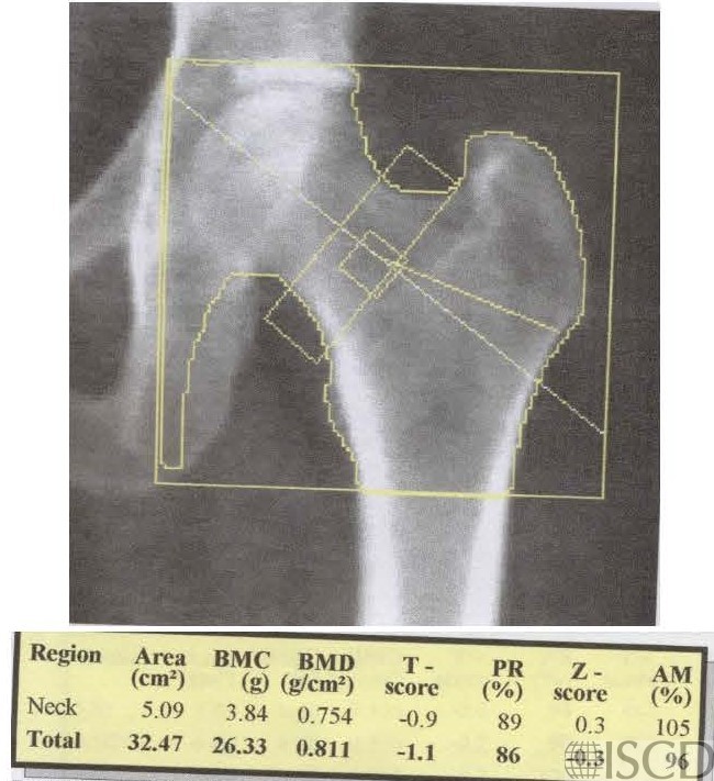
This is the baseline Hologic left hip scan.
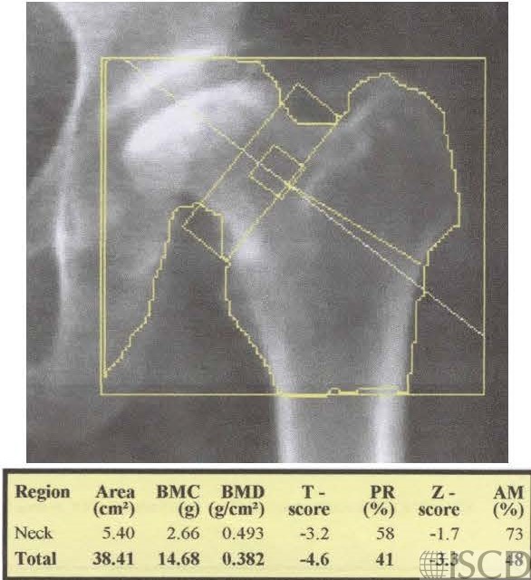
This is the follow-up left Hologic hip scan 8 years later after an episode of thyroid storm and chronic steroid use for rheumatoid arthritis.
This is the accompanying hip radiograph showing avascular necrosis of the left femoral head.

These images show a baseline DXA and a follow-up DXA 8 years later in a patient who had intervening thyroid storm and was placed on chronic steroids for rheumatoid arthritis. The follow-up image shows avascular necrosis with collapse and remodeling. There is a significant decrease in bone mineral density in the 8 years after starting steroids and thyroid storm. It was not possible to rotate the hip internally to the same extent as on the baseline scan because of the change in hip anatomy.
Sarah L Morgan, MD, RD, CCD, The University of Alabama at Birmingham
• Mitchell, D.G., et al., Femoral head avascular necrosis: correlation of MR imaging, radiographic staging, radionuclide imaging, and clinical findings. Radiology, 1987. 162(3): p. 709-15.