Gluteal Implant

There is a gluteal implant overlying the left proximal femur in this Hologic left hip scan. The left proximal femur would not be useful for analysis because of the overlying artifact. A right hip scan should be completed and analyzed.
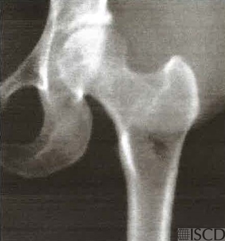
This Hologic left proximal femur scan shows an overlying gluteal implant.
Sarah L Morgan, MD, RD, CCD, The University of Alabama at Birmingham
• Martineau, P., S. Bazarjani, and L.S. Zuckier, Artifacts and Incidental Findings Encountered on Dual-Energy X-Ray Absorptiometry: Atlas and Analysis. Semin Nucl Med, 2015. 45(5): p. 458-69.
• Bazzocchi, A., et al., Incidental findings with dual-energy X-ray absorptiometry: spectrum of possible diagnoses. Calcif Tissue Int, 2012. 91(2): p. 149-56.
• Bojinca, V., D. Opris, and M. Bonjinca, Artifacts and pitfalls in DXA scan images and interpretation. J Clin Densitom, 2012. 15(4): p. 486-487.
• Friedman, G., et al., Case report: Effect of silicone buttock implant on DXA scan evaluation. . J Clin Densitom, 2010. 13(1): p. 135.
• Hassan, A., et al., Effect of silicone gluteal implant on bone mineral density evaluation by DXA scan. . J Clin Densitom, 2011. 15(1): p. 124-128.
• Hauache, O.M., et al., Increased hip bone mineral density in a woman with gluteal silicon implant. J Clin Densitom, 2000. 3(4): p. 391-3.
• Rivera, M., P. Humeres, and P. Gonzalez, Increased bone mineral density in dual x-ray densitometry due to gluteal implants. Clin Nucl Med, 1999. 24(1): p. 51-3.