Breast Implants

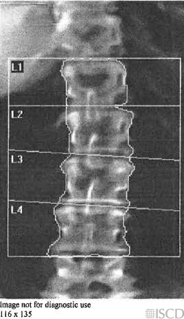
This Hologic lumbar spine image shows a breast implant lateral to L1 on the right.
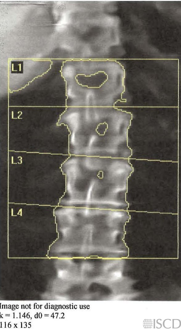
This image shows the corresponding undo view which demonstrates that the breast implant would be removed from the soft tissue baseline.
This is the corresponding chest x-ray for the DXA scan showing the breast implants.
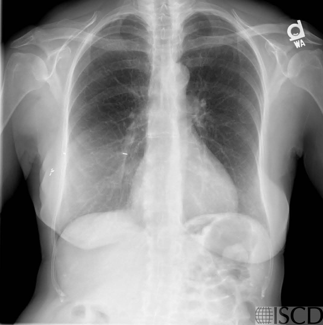
This Hologic DXA scan shows a breast implant lateral to L1 on the right. The corresponding undo view shows that the breast implant would be removed from the soft tissue baseline. The correspond chest radiograph shows the implants.
Sarah L Morgan, MD, RD, CCD, The University of Alabama at Birmingham
References